Human Anatomy and Physiology - N. I. Fedyukovich 2003
Internal Organs
Digestive System
Stomach
The Stomach (ventriculus, gaster) is an expanded part of the digestive tract that serves as a reservoir for food and is located between the Esophagus and the duodenum.
The stomach is divided into the anterior and posterior walls, the lesser and greater curvatures, the cardiac part, the fundus (vault), the body, and the pyloric part (Fig. 76).
Class="center">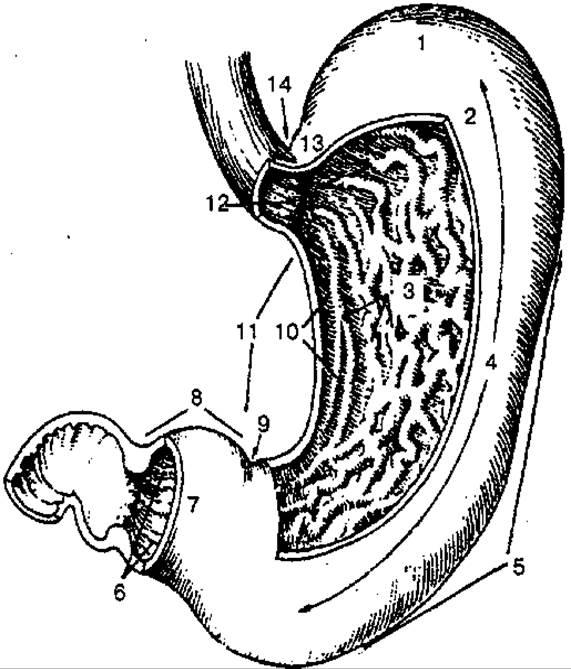
Fig. 76. The stomach (opened):
1 — Cytology/practical/108.html">Fundus of the stomach; 2 — anterior wall; 3 — gastric folds; 4 — body of the stomach; 5 — greater curvature of the stomach; 6 — pyloric canal; 7 — pyloric antrum; 8 — pyloric part; 9 — angular notch; 10 — gastric canal; 11 — lesser curvature of the stomach; 12 — cardiac orifice; 13 — cardiac part of the stomach; 14 — cardiac notch
The size of the stomach varies greatly depending on body build and the degree of fullness of the organ. When moderately filled, the stomach has a length of 24—26 cm, and 18—20 cm when empty. The capacity of an adult stomach averages 3 L (1.5—4.0 L).
The stomach wall consists of the mucosa, submucosa, muscularis, and serosa.
The gastric mucosa is lined with a simple columnar epithelium and forms numerous folds running in different directions: longitudinal along the lesser curvature, and transverse, oblique, and longitudinal in the fundus and body. At the junction of the stomach and the duodenum, There is a circular fold—the pyloric valve—which, when the pyloric sphincter contracts, separates the gastric cavity from the duodenum. The mucosa features small elevations known as gastric areas. On the surface of these areas are small depressions (gastric pits) that represent the openings of the gastric glands, which secrete gastric juice for the chemical Digestion of food.
The gastric submucosa is well-developed and contains dense vascular and nerve plexuses.
The muscularis of the stomach (Fig. 77) consists of an inner oblique layer of Muscle fibers, a middle circular layer, and an outer longitudinal layer of smooth muscle fibers. In the pyloric region, the circular layer is more developed than the longitudinal layer, forming the pyloric sphincter around the outlet.

Fig. 77. Muscularis of the stomach:
1,8 — longitudinal layer; 2 — oblique fibers; 3, 4 — circular layer; 5 — pylorus; 6 — pyloric orifice; 7 — pyloric sphincter; 9 — muscularis
The stomach is located in the upper Abdominal cavity, beneath the Diaphragm and the Liver. Three-quarters of it lies in the left hypochondrium, and one-quarter in the epigastric region. The cardiac orifice is located at the level of the bodies of the X—XI thoracic vertebrae, while the pyloric orifice lies at the right margin of the XII thoracic and I lumbar vertebrae.
The longitudinal axis of the stomach runs obliquely from above downward, from left to right, and from back to front. The anterior surface of the stomach, in the cardiac part, fundus, and body, is in contact with the diaphragm, and in the region of the lesser curvature, with the left lobe of the visceral surface of the liver. A small portion of the body of the stomach lies directly adjacent to the anterior abdominal wall.
Along the greater curvature, the posterior surface of the stomach is in contact with the transverse colon, and in the region of the fundus, with the Spleen.
Behind the stomach lies a slit-like space—the omental bursa—which separates it from the Organs situated on the posterior abdominal wall: the left Kidney, Adrenal gland, and Pancreas. The relatively stable position of the stomach is maintained by its connection to surrounding organs via the hepatogastric, gastrocolic, and gastrosplenic ligaments.
Last update: 08/08/2026
Editorial and Educational Adaptation: This material has been compiled based on the primary/original source text. The project team performed an editorial review, corrected technical inaccuracies, structured sections, and adapted the content for an educational format.
What was processed:
- elimination of formatting defects (OCR errors, structural breaks, corrupted characters);
- editorial organization of content;
- standardization of terminology in accordance with academic sources;
- verification of factual statements against the original source text.
All mentions of the author, publication year, and origin of the primary text have been preserved in accordance with the source.