Human Anatomy, Part 1 - K. A. Dyubenko, A. K. Kolomiysev, Yu. B. Chaykovsky 2002
Special Section
Splanchnology, splanchnologia [the study of viscera] — Development of the digestive and respiratory systems
Development of the respiratory organs
This process begins at the end of the fourth week of Embryogenesis. They originate in the lower pharyngeal gut, just below the last pharyngeal pouch. Here, on the ventral surface of the primitive gut, an outpocketing appears—the laryngotracheal groove. As it separates from the gut, it forms a tube that communicates with the posterior Pharynx. This tube represents the primordial rudiment of the Larynx and Trachea. The larynx develops from the cranial portion of the laryngotracheal bud, whereas the more caudally situated part of the tube gives rise to the trachea.
Already in the fourth week, a dilatation forms at the caudal end of this rudiment and divides into two outpocketings—the right and left lung buds (Fig. 279), which serve as the precursors of the main Bronchi and lung lobes.
Class="center">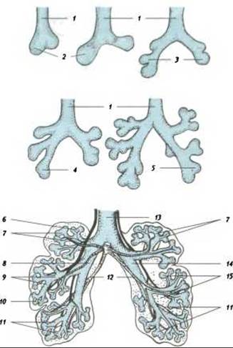
Fig. 279. Development of the bronchi and Lungs (after Patten):
1 - trachea; 2 - pulmonary sac; 3 - primary bronchus; 5 - left main bronchus; 6 - apical bronchus; 7 - superior lobe of the lung; 8 - right bronchus; 9 - middle lobe of the lung; 10 - primordium of the visceral pleural layer; 11 - inferior lobe of the lung; 12 - Pulmonary Veins; 13 - bifurcation; 14 - left bronchus; 15 - mesenchyme of the pulmonary stroma
The epithelium of both the respiratory tract and the respiratory portions of the lungs is derived from the foregut lining. Cartilage, Connective Tissue, and Muscle tissue are formed from the mesenchyme.
The right lung bud (which is larger) grows dorsocaudally, while the left one (which is smaller) grows in a more lateral direction. Subsequently, the rudiment of the right main bronchus divides into three segmental bronchi, and the left into two, meaning that the right lung consists of three lobes and the left of two. With further growth, the bronchi undergo dichotomous branching, resulting in a continuous increase in the number of bronchial tree branches. The formation of the bronchi is generally completed by the seventh month of embryogenesis, though it continues up until birth and even During the first postnatal months.
Shortly before birth, the concentration of surface-active agents increases in the lungs; these substances reduce the surface tension of the fluid layer lining the inner surface of the alveoli and prevent their collapse. The production of these substances, collectively known as surfactant, is regulated by Hormones secreted by the adrenal cortex.
The fetal lungs are filled with fluid secreted by the lung Tissues. This fluid is partially expelled during delivery by mechanical compression and partially absorbed by lymphatic and Blood microvessels.
The Pleura develops from the ventral mesoderm: specifically, the visceral pleura arises from its visceral layer, and the parietal pleura from its parietal layer.
Last update: 08/08/2026
Editorial and Educational Adaptation: This material has been compiled based on the primary/original source text. The project team performed an editorial review, corrected technical inaccuracies, structured sections, and adapted the content for an educational format.
What was processed:
- elimination of formatting defects (OCR errors, structural breaks, corrupted characters);
- editorial organization of content;
- standardization of terminology in accordance with academic sources;
- verification of factual statements against the original source text.
All mentions of the author, publication year, and origin of the primary text have been preserved in accordance with the source.