Human Anatomy Part 1 - K. A. Dyubenko, A. K. Kolomiytsev, Yu. B. Chaykovsky 2002
Special Part
Splanchnology, splanchnologia [the study of viscera] - Digestive system, systema digestorium
Esophagus
The oesophagus (Esophagus) is a muscular tubular organ that forms part of the gastrointestinal tract (Fig. 222). It serves to convey food from the Pharynx to The Stomach. The oesophagus begins at the level of the sixth cervical vertebra (CVI), and its inferior margin lies at the level of the eleventh thoracic vertebra (TXI). In adults, the length of the oesophagus ranges from 25 to 30 cm, with the distance from the dental arch to the stomach being 40-42 cm—a crucial consideration when performing sounding Procedures.
Topography of the oesophagus. Based on its anatomical Location, the oesophagus is divided into the following parts (see Fig. 222):
- cervical part, pars cervicalis;
- thoracic part, pars thoracica;
- abdominal part, pars abdominalis.
The cervical part, pars cervicalis, extends from the sixth cervical vertebra, posterior to the cricoid Cartilage, down to the third thoracic vertebra; its length is 5-6 cm. Anteriorly, the oesophagus lies adjacent to the Trachea. The recurrent laryngeal nerves run within the grooves between the oesophagus and trachea, which must be taken into account during surgical interventions in this region. The lateral surfaces of the oesophagus are in contact with The Thyroid Gland. Inferiorly, on the left side, the Thoracic duct abuts the oesophagus. Posterior to the oesophagus lies the retrooesophageal space, which is a continuation of the retropharyngeal space and communicates with the posterior Mediastinum.
The thoracic part, pars thoracica, is located in the posterior mediastinum, spanning from the second thoracic vertebra to the oesophageal hiatus of the Diaphragm. Its length is 15-18 cm. Anteriorly, the thoracic part is related to the left recurrent laryngeal nerve, Branches of the left Vagus nerve, the oesophageal nerve plexus, the left common carotid artery, the arterial bifurcation, and the left principal bronchus. To the left of the oesophagus lie the left Subclavian Artery, the left vagus nerve, the thoracic duct, the aortic arch, and the Thoracic Aorta. To the right are branches of the vagus nerve and the azygos vein. Posteriorly, the thoracic segment of the oesophagus is related to the longus colli Muscles, the cervical vertebrae, the thoracic duct, the hemiazygos vein, and the thoracic aorta.
Class="center">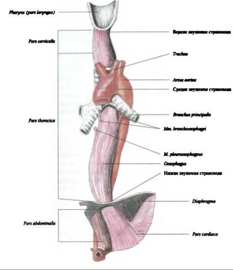
Fig. 222. Oesophagus, oesophagus (anterior view)
The abdominal part, pars abdominalis, extends from the oesophageal hiatus of the diaphragm to the cardiac orifice of the stomach, measuring 2 to 4 cm in length. In this region, the oesophagus is covered by Peritoneum anteriorly and laterally. Anteriorly, it is related to the left lobe of the Liver, and to the left, to the superior pole of the Spleen. Initially, in the cervical region, the oesophagus runs to the left of the midline down to the sixth cervical vertebra. From the level of the fifth thoracic vertebra, it courses along the midline, then curves to the right at the eighth thoracic vertebra, before shifting to the left once again.
The oesophagus presents three anatomical constrictions:
- the pharyngeal constriction, located at the pharyngooesophageal junction at the level of the sixth cervical vertebra (CVI);
- the bronchial constriction, located where it is crossed by the left principal bronchus at the level of the fourth to sixth thoracic vertebrae (TIV-TVI);
- the diaphragmatic constriction, located at the oesophageal hiatus of the diaphragm at the level of the tenth to eleventh thoracic vertebrae (TX-TXI).
Physiological constrictions occur exclusively in the living subject. They are situated at the point where the oesophagus is crossed by the aorta (aortic constriction) and at the junction of the oesophagus with the cardiac part of the stomach (cardiac constriction). Foreign bodies may become lodged at these sites, a factor of significant clinical importance.
Last update: 08/08/2026
Editorial and Educational Adaptation: This material has been compiled based on the primary/original source text. The project team performed an editorial review, corrected technical inaccuracies, structured sections, and adapted the content for an educational format.
What was processed:
- elimination of formatting defects (OCR errors, structural breaks, corrupted characters);
- editorial organization of content;
- standardization of terminology in accordance with academic sources;
- verification of factual statements against the original source text.
All mentions of the author, publication year, and origin of the primary text have been preserved in accordance with the source.