Human Anatomy Part 1 - K. A. Dyubenko, A. K. Kolomiytsev, Yu. B. Chaikovsky 2002
Special Part
Myology, myologia [the study of muscles] - Muscles and fasciae of the lower limb
Muscles of the lower limb, musculi membri inferioris - Pelvic muscles - Internal group
1. Iliopsoas Muscle, m. iliopsoas — formed by the confluence of the fibers of two Muscles: the m. iliacus and m. psoas major (Fig. 171). It emerges from the pelvic cavity through the muscular lacuna and attaches to the lesser trochanter of the Femur. The iliopsoas muscle participates in forming the posterior wall of the Abdominal cavity AND plays an important role in developing lumbar lordosis.
Class="center">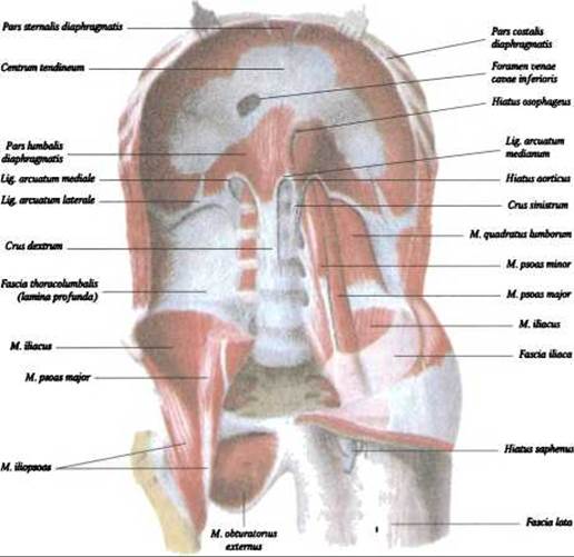
Fig. 171. Muscles of the pelvis (internal group) and Diaphragm (according to R. D. Sinelnikov)
2. Psoas major muscle, m. ilioрsоаs major (see Fig. 171) — originates from the lateral surface of the XII thoracic and I–IV lumbar vertebrae. Deep muscle fibers originate from the transverse processes of all lumbar vertebrae, course inferiorly, and together with the m. iliacus form the iliopsoas muscle, m. iliopsoas.
Function: flexes the thigh at the hip joint, laterally rotates it (supination), and when the thigh is fixed, bends the trunk forward. When standing on one leg, this muscle not only flexes the pelvis but also rotates it around a vertical axis passing through the hip joint.
Blood supply: aa. iliolumbalis, circumflexa iliasus profunda.
Innervation: rr. musculares plexus lumbalis (I 1-І II).
3. Psoas minor muscle, m. psoas minor (see Fig. 171) — inconstant, located on the anterior surface of the m. psoas minor, originates from the lateral surface of the body of the XII thoracic and I lumbar vertebra, and attaches to the pubic crest and eminentia iоpeсtinea.
Function: tensions the fascia iliaca.
Blood supply: aa. lumbales.
Innervation: rr. musculares plexus lumbalis (I 1-І II).
4. Iliacus muscle, m. iliacus (see Fig. 171) — fills the fossa iliaca, originates from its walls, with fibers converging in a fan-like manner toward the pelvic brim, and together with the m. psoas major forms the iliopsoas muscle, m. iliopsoas.
5. Piriformis muscle, m. piriformis — triangular in shape, originates from the anterior surface of the sacrum near the foramina sacralis (II–V) (Figs. 172, 173), courses laterally and inferiorly, leaves the lesser pelvic cavity through the greater sciatic foramen (foramen ischiadicum majus), and attaches to the apex of the greater trochanter (trochanter major) (see Fig. 172).
Function: laterally rotates the thigh (supinates) and also participates in its abduction.
Blood supply: aa. glutea superior et inferior.
Innervation: rr. musculares plexus sacralis (5 1-5 II (5 III)).
6. Obturator internus muscle, m. obturator internus — located on the inner surface of the lateral wall of the lesser pelvis (see Fig. 173). It originates from the inner surface of the os coxae around the obturator membrane (membrana obturatoria) and from its inner surface. The muscle passes through the lesser sciatic foramen (foramen ischiadicum minus), enters the gluteal region, and attaches to the trochanteric fossa (fossa trochanterica). The superior and inferior gemelli muscles attach to the tendon of this muscle.
Function: the function of the obturator internus and gemelli muscles is to abduct the thigh, and when standing on one leg, these muscles prevent the pelvis from tilting to the opposite side.
Blood supply: aa. gfutea inferior, obturatoria, pudenda interna.
Innervation: rr. musculares plexus sacralis (I IV-5 II (5 III)).
Last update: 08/08/2026
Editorial and Educational Adaptation: This material has been compiled based on the primary/original source text. The project team performed an editorial review, corrected technical inaccuracies, structured sections, and adapted the content for an educational format.
What was processed:
- elimination of formatting defects (OCR errors, structural breaks, corrupted characters);
- editorial organization of content;
- standardization of terminology in accordance with academic sources;
- verification of factual statements against the original source text.
All mentions of the author, publication year, and origin of the primary text have been preserved in accordance with the source.