Human Anatomy, Part 1 - K. A. Dyubenko, A. K. Kolomiytsev, Yu. B. Chaykovsky 2002
Special Part
Arthrology, artrologia [the study of bone joints] — Classification of joints and their general characteristics
Joints of the skull
The cranial bones are connected to one another by cranial sutures, suturae cranii. These sutures are named after the bones they connect, as well as their position and Morphology (serrate, squamous, or plane). The suture between the frontal and parietal bones is called the coronal suture, sutura coronalis; between the parietal bones, the sagittal suture, sutura sagittalis; and between the parietal and occipital bones, the lambdoid suture, sutura lambdoidea. The BONES OF THE facial Skeleton are connected by plane sutures, suturae planae. The bones of the Skull base are joined by fibrous Cartilage located in the gaps between individual bones. This type of articulation is known as a synchondrosis. An example is the junction between the bodies of the occipital and sphenoid bones, synchondrosis sphenooccipitalis.
The temporomandibular joint, articulatio temporomandibularis, is a combined articulation that, based on its range of motion, can be classified as ginglymoid (hinge-like). It is formed by the HEAD of the Mandible, caput mandibulare, and the mandibular fossa, fossa mandibularis, of the Temporal bone (Fig. 86). The mandibular head has an ellipsoidal shape oriented transversely. The mandibular fossa is larger than the articular head, and congruency between them is achieved through the articular disc, discus articularis.
The temporomandibular joint features an articular tubercle bounding the fossa anteriorly, as well as an articular disc, discus articularis. The disc is lens-shaped and biconcave. It divides the joint cavity into two isolated compartments, serving to harmonize the articular surfaces and cushion chewing forces.
The capsule of the temporomandibular joint attaches along the margin of the fossa mandibularis; posteriorly, along the petrotympanic fissure, fissura petrotympanica; and medially, along the tympanosquamous (tympanic) suture, sutura tympanica. On the mandible, the capsule attaches to the neck of the articular process.
The ligaments of the temporomandibular joint are divided into two groups:
1. Intracapsular ligaments:
- anterior and posterior discotemporal ligaments, ligg. discotemporales anterior et posterior,
- lateral and medial discomandibular ligaments, ligg. discomandibulares laterale et mediale.
2. Extracapsular ligaments:
- lateral ligament, lig. laterale;
- sphenomandibular ligament, lig. sphenomandibulare;
- stylomandibular ligament, lig. stylomandibulare.
The following movements occur in the temporomandibular joint: elevation and depression of the mandible, as well as its protrusion (forward movement) and lateral excursions.
Blood supply is provided by the maxillary artery, a. maxillaris. Venous blood drains into the venous plexus of the joint and the retromandibular vein.
Lymphatic drainage is directed to the parotid Lymph Nodes, nodi lymphatici parotidei, and deep cervical nodes.
Innervation of the joint is supplied by the auriculotemporal nerve, n. auriculotemporalis, a branch of the mandibular division of the Trigeminal nerve.
Class="center">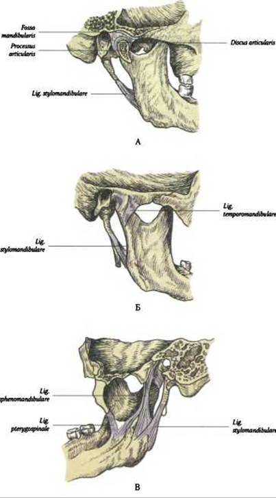
Fig. 86. Structure and ligaments of the temporomandibular joint:
A - sagittal section; B - external surface; C - internal surface
Last update: 08/08/2026
Editorial and Educational Adaptation: This material has been compiled based on the primary/original source text. The project team performed an editorial review, corrected technical inaccuracies, structured sections, and adapted the content for an educational format.
What was processed:
- elimination of formatting defects (OCR errors, structural breaks, corrupted characters);
- editorial organization of content;
- standardization of terminology in accordance with academic sources;
- verification of factual statements against the original source text.
All mentions of the author, publication year, and origin of the primary text have been preserved in accordance with the source.