Protein Structure and Function: Application of Bioinformatics Methods - John Rigden 2014
Protein Dynamics: From Structure to Function
Methods for Predicting Functional Modes
CONCOORD
The CONCOORD program (de Groot et al. 1997) employs a geometric approach to predict protein flexibility. The three-dimensional Structure of a protein is determined by various interactions, such as covalent bonds, Hydrogen Bonds, and non-polar interactions. Most of these interactions remain unchanged during functionally significant conformational transitions. This observation forms the core of the CONCOORD method: starting from the initial structure data, alternative structures are generated in which the vast majority of interactions are preserved. To achieve this, the first step of the CONCOORD calculation analyzes the interactions in the starting structure and derives geometric constraints from them—primarily upper and lower bounds on atomic distances, as well as angle constraints and data on planar and chiral groups. This geometric Description of the structure can be thought of as a blueprint for protein assembly. In the second step, beginning from a randomized atomic arrangement, the structure is iteratively rebuilt based on the previously established blueprint, typically repeating this process several times. Because each run starts from random atomic positions, unlike Molecular Dynamics (MD), this method does not suffer from sampling limitations and yields an ensemble that covers the entire conformational space accessible under the specified constraints. However, the method provides no information regarding the pathway between substates, the transition time, or the transition energy (Fig. 9.12).
Class="center">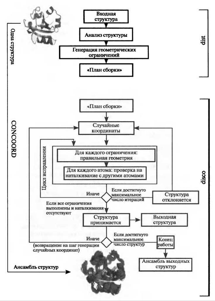
Fig. 9.11. Schematic representation of the CONCOORD method for generating structural ensembles from a single starting structure. In the first step (the dist program), the initial structure is analyzed and converted into a geometric description of the protein. In the second step (the disco program), starting from random coordinates, the structure is reconstructed based on the previously defined constraints.
9.4.3.1. Applications
CONCOORD and its recently developed extension tCONCOORD (Seeliger et al. 2007) have been applied to investigate various Proteins. Adenylate kinase exhibits a pronounced domain-closing motion upon binding a substrate (ATP/AMP) or an inhibitor (see top of Fig. 9.13), with a Ca RMSD of 7.6 Å between the Ligand-bound and ligand-free Conformations. Two calculations were performed using tCONCOORD, both starting from the closed conformation (PDB code 1AKE) as the initial state. In one of the calculations, the ligand (Ap5A) was removed. The bottom of Fig. 9.13 shows the results of a Principal Component Analysis (PCA) performed on experimental structures. The first principal component (abscissa) corresponds to the domain-opening motion indicated by the arrow in Fig. 9.13 (bottom). Each data point on the plot represents a single structure. Red points represent the ensemble generated from the ligand-free closed conformation of adenylate kinase. Green points represent the ensemble generated from the ligand-bound conformation. Although the calculation with the inhibitor sampled predominantly closed conformations in the vicinity of the ligand-bound structure, the calculation without the ligand successfully sampled both closed and open conformations, successfully approaching the experimentally determined open structures with RMSDs of 2.4, 2.6, and 3.1 Å for 1DVR, 1AK2, and 4AKE, respectively. Structure-based drug design often faces the inverse problem: predicting the ligand-bound receptor structure starting from the free receptor. A calculation starting from the open conformation (4AKE) successfully generated structures approaching the closed conformation with RMSDs of 2.5, 2.9, and 3.3 Å for 1DVR, 1AK2, and 4AKE, respectively. Thus, the functional domain-opening motion was successfully predicted in both cases—whether starting from the closed, ligand-bound conformation or the open, ligand-free conformation.

Fig. 9.12. Comparison of the sampling properties of molecular dynamics and the CONCOORD method on a hypothetical free-energy landscape. The MD trajectory (left) wanders across the surface, thereby providing information about the time and pathway between conformational substates. CONCOORD (non-deterministically) hops across the surface, thereby achieving a more thorough sampling of the conformational space.

Fig. 9.13. (For the color version of this figure, please see the color insert.) Top: Superposition of X-ray crystal structures of adenylate kinase. Bottom: Principal component analysis. Two structural ensembles generated by tCONCOORD are projected onto the first two eigenvectors derived from the PCA of the X-ray structure ensemble. The ensemble shown in red was generated from the closed conformation (1AKE) with the inhibitor removed. This generated ensemble samples both closed and open conformations. The ensemble shown in green was also generated from the closed conformation (1AKE), but in the presence of the inhibitor. This ensemble samples exclusively closed conformations in the vicinity of the initial ligand-bound structure.
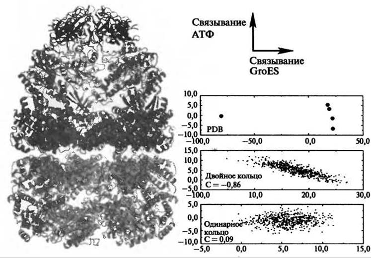
Fig. 9.14. Asymmetric GroEL-GroES complex (left) along with the results of CONCOORD calculations (right). The GroEL-GroES complex consists of two GroES co-chaperonins (shown in black), a GroEL trans-ring bound to GroES (shown in dark gray), and a cis-ring (shown in light gray). Principal component analysis revealed two primary structural transitions for the GroEL ring: upon nucleotide binding (vertical axis on the right panel) and upon GroES binding (horizontal axis), respectively. In the double-ring calculations—unlike the single-ring ones—these modes were found to be coupled, suggesting a direct link between intra-ring and inter-ring cooperativity.
Due to its computational efficiency, CONCOORD is well-suited for identifying functionally relevant modes of flexibility in molecular systems that exceed the size limitations of other atomic-detail Methods, such as molecular dynamics. Applying CONCOORD to the GroEL-GroES chaperonin complex, comprising over 8,000 residues, uncovered a novel form of coupling between intra-ring and inter-ring cooperativity (de Groot et al. 1999). Each GroEL ring exhibited two major collective motion modes: a primary conformational transition upon binding the GroES co-chaperonin, and a secondary transition upon ATP binding (Fig. 9.14, top right). CONCOORD calculations on a single GroEL ring alone failed to show any coupling between these modes, whereas simulations of the double-ring system revealed a clear correlation between them, thereby explaining how nucleotide binding modulates affinity for GroES in the double ring, but not in the single ring.
Last update: 06/08/2026
Editorial and Educational Adaptation: This material has been compiled based on the primary/original source text. The project team performed an editorial review, corrected technical inaccuracies, structured sections, and adapted the content for an educational format.
What was processed:
- elimination of formatting defects (OCR errors, structural breaks, corrupted characters);
- editorial organization of content;
- standardization of terminology in accordance with academic sources;
- verification of factual statements against the original source text.
All mentions of the author, publication year, and origin of the primary text have been preserved in accordance with the source.