Biotechnology - Yu.O. Sazykin 2006
Applied Biotechnology
Challenges in the discovery, development, and application of antibiotics in medical practice
Mechanisms of antibiotic action — Inhibitors of bacterial cell wall synthesis
Inhibition occurs due to the selective suppression of certain Enzymes involved in the multi-step Synthesis of the primary Cell wall polymer, peptidoglycan. Peptidoglycan synthesis inhibitors include Cephalosporins and other beta-lactam compounds.
As the name suggests, peptidoglycan consists of fundamentally distinct components. Its long, parallel glycan strands are built from alternating residues of two sugars: N-acetylglucosamine and muramic acid. Muramic acid (derived from the Greek μupuζ, meaning wall, as it was first discovered in the Introduction/37.html">Bacterial cell wall) is an ether of N-acetylglucosamine and lactic acid. The peptide portion of the polymer comprises short peptide chains (three to five amino acid residues) branching off from the muramic acid residues within the glycan.
The peptide chains branching off from the parallel glycan strands in this manner are linked together by peptide bonds. Transverse peptide "bridges" form between the parallel glycan strands, creating a complete, integrated peptidoglycan that envelops the entire bacterial cell in a rigid "mesh" (Fig. 14).
Gram-positive Bacteria possess a thick peptidoglycan layer, whereas in Gram-negative bacteria, it is significantly thinner.
Beta-lactam Antibiotics (without exception) inhibit the enzyme-catalyzed closure of peptide chains into peptide bridges during peptidoglycan Biosynthesis. The glycan strands remain unconnected, preventing The formation of the continuous, cell-encompassing peptidoglycan network. Consequently, peptidoglycan loses the properties essential for the bacterial cell's survival. Bacterial Cells continue to divide, grow, and divide again—processes that require ongoing peptidoglycan synthesis and the insertion of newly synthesized fragments into the polymer, which is cleaved by specific peptidoglycan Hydrolases for this purpose. Under normal conditions, The Cell maintains a balance between the enzymes that hydrolyze peptidoglycan and those that synthesize it.
Class="center">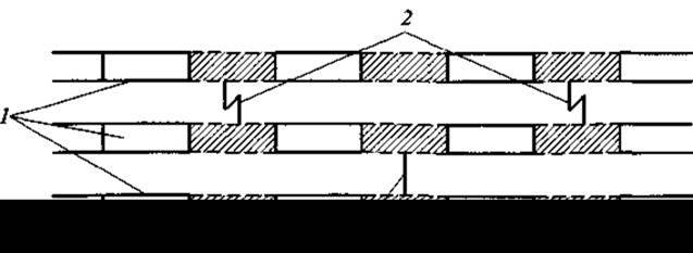
Fig. 14. Fragment of the peptidoglycan Structure:
1 — glycan; 2 — unlinked peptide chains; 3 — peptide bridges
In the presence of beta-lactam antibiotics, this balance is disrupted. Peptidoglycan synthesis ceases, but its Enzymatic Hydrolysis continues and even accelerates because The cell wall loses certain components that normally constrain hydrolase activity through reversible blockage. Both the glycan strands and peptide bridges undergo hydrolysis. The Mechanism of the antibiotic activity of beta-lactams is linked to target enzymes that have no analogs in animal cells. Animal cells lack a rigid cell wall, peptidoglycan, and, consequently, the enzymes responsible for its synthesis. Thus, beta-lactams are non-toxic to The Human Body; if certain beta-lactams exhibit toxicity or allergenicity under specific conditions, these effects are entirely unrelated to the mechanism of their antibacterial action.
Last update: 06/08/2026
Editorial and Educational Adaptation: This material has been compiled based on the primary/original source text. The project team performed an editorial review, corrected technical inaccuracies, structured sections, and adapted the content for an educational format.
What was processed:
- elimination of formatting defects (OCR errors, structural breaks, corrupted characters);
- editorial organization of content;
- standardization of terminology in accordance with academic sources;
- verification of factual statements against the original source text.
All mentions of the author, publication year, and origin of the primary text have been preserved in accordance with the source.