Protein Chemistry - Part 1 - General Protein Chemistry - Ashmarin I. P. 1968
Isolation and Purification of Proteins
Separation of Protein Mixtures
Preparative Electrophoresis
The Description of the electrochemical Properties of Proteins and the corresponding Analytical Methods is the subject of subsequent sections of this manual, which also outlines The Theory of Protein Electrophoresis. Here, we shall only note that proteins differ significantly from one another in their mobility in an electric field. If the pH of the solution corresponds to the isoelectric point of a given protein, its net charge is zero, and the electric field will not affect its movement. As the pH decreases, the net charge becomes positive, and the protein migrates toward the cathode. At a pH above the isoelectric point, the protein moves toward the anode. The greater the difference between the solution pH and the isoelectric point of the protein, the higher the migration velocity. However, differences in the electrophoretic mobility of proteins depend on more than just the mismatch of their isoelectric points. Even assuming identical isoelectric points for two proteins, their velocities in an electric field at the same pH value will still differ. This is due to variations in the number and nature of polar groups, which alter the charge to differing degrees when the pH deviates from the isoelectric point. In addition, the size and shape of the protein molecule influence its migration rate. As a result, protein fractionation based on differential electrophoretic mobility proves to be one of the most powerful methods.
Here, we will consider only those variants of electrophoresis that are suitable for separating substantial quantities of protein mixtures while isolating individual fractions. The application of free solution electrophoresis without any supporting media for this purpose is hindered by the same circumstances that complicate preparative ultracentrifugation. When attempting to isolate the zone of the solution containing a particular fraction, liquid currents easily arise, blurring the boundaries and mixing the contents of different zones. Numerous methodological techniques have been proposed to overcome this obstacle. For instance, at a certain stage of the process, part of the cuvette was cut off by an advancing Glass plate. Porous partitions were also used; while they did not impede the movement of protein particles, they prevented mixing when a portion of the cuvette contents was withdrawn. These and other similar techniques have not become widely adopted, primarily because they allow only the fractions occupying the extreme positions in the cuvette to be obtained in pure form. Furthermore, the throughput of these methods is relatively low, and the required equipment is rather expensive and complex to operate.
Only so-called electrodecantation has achieved somewhat wider use. This technique is implemented as follows: if electrophoresis is carried out in a Cell bounded on the electrode sides by semipermeable membranes, protein particles migrating toward the electrodes concentrate near them. The resulting layer of concentrated protein solution gradually "slides" down to the bottom of the cuvette, as shown in Fig. 4. By the end of the process, only the protein whose isoelectric point coincides with the pH of the solution—and which is therefore immobile in the electric field—remains in the upper part of the cuvette. Instruments have been developed in which this process is continuous, such that fresh portions of the protein mixture are gradually fed into the cuvette, while the purified protein solution is suctioned off from the upper section. The cuvettes in such devices are made as narrow as possible to reduce the distance that the removed proteins must travel to reach the membrane. Instruments consisting of consecutively connected batteries of such cuvettes have been described and are recommended, in particular, for the industrial purification of certain Serum proteins.
However, electrophoresis using various supporting media—known as zonal electrophoresis, i.e., electrophoresis in solutions impregnating a solid porous or powdered carrier such as filter paper, starch, Cellulose, or glass powder—has proven more practical and effective. Even more efficient, though less convenient for preparative purposes, is electrophoretic Separation in gels, such as Agar-agar, starch, or polyacrylamide gels.
Class="center">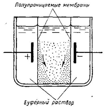
Fig. 4. Schematic diagram of an electrodecantation device. Explanations in the text.
Zonal electrophoresis on porous or powdered carriers does not fundamentally differ from free electrophoresis. In this case as well, the fractionation of proteins is determined primarily by differences in their mobility in an electric field. However, the isolation of individual fractions is greatly facilitated. The schematic diagram of a device for zonal electrophoresis is shown in Fig. 5.
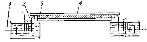
Fig. 5. Schematic diagram of an apparatus for zonal electrophoresis on a powdered carrier:
1 — electrodes, 2 — filter paper bridges, 3 — starch block, 4 — glass plate.
The carrier, impregnated with a buffer solution, is placed into an elongated cuvette and compacted to form a moist block. Then, a small section of the block is replaced with carrier material impregnated with the protein solution to be fractionated. A potential difference is applied across the ends of the block, causing the proteins to migrate from the point of application toward their respective electrodes. Characteristically, under these conditions, the path a protein can travel is tens of times greater than the size of the application zone. This is one of the advantages of zonal electrophoresis over free electrophoresis, where the maximum migration distance of the particles barely exceeds the width of the initial zone. This makes it possible to resolve practically all fractions from one another, rather than just the extreme ones. After the individual fractions have migrated sufficiently apart, the block is cut into small sections from which the respective fractions can then be extracted. Fraction mixing is practically absent in this process. Preparative electrophoresis on a starch block has become particularly widespread. Protein sorption on starch particles is usually negligible, ensuring good protein recovery from the corresponding section. Protein sorption on the other carriers listed above is generally more significant, which complicates their widespread application for preparative purposes. Filter paper electrophoresis has proved very convenient as an analytical method.
Continuous-flow apparatus based on the same principle have been described in the literature. They utilize flat chambers filled with a relatively coarse-grained carrier (glass powder) or a vertically positioned sheet of filter paper. The protein solution is continuously fed onto one of the sections of the upper edge of the block or paper sheet. Moving toward the respective electrodes while simultaneously descending with the liquid flow, the protein particles reach the lower edge at different locations. The dripping liquid is collected at multiple points, making it possible to isolate the fractions.
Regarding gel electrophoresis, its fundamental differences from electrophoresis in powdered or porous Supports should be emphasized. Gels serve not merely as a supporting medium for the solution; they also function as molecular sieves, altering the migration velocity of protein molecules depending on their size and shape. Thus, two Protein Separation Methods are combined: electrophoretic separation and molecular sieving. As a result, the resolving power of gel electrophoresis is the highest. The instrumentation for this method has no special features. A minor drawback of gel electrophoresis is the relative difficulty of extracting fractions from the gel segments.
In all variants of preparative electrophoresis, an important task is to protect the protein solution from decomposition products of the solvent components generated at the electrodes, as well as from the heating caused by high electric voltages. Therefore, the electrodes are placed in special compartments that communicate with the protein migration zone via a system of electrolytic bridges and intermediate vessels containing Buffer solutions (see Fig. 5). Special cooling devices are also frequently employed.
Last update: 06/08/2026
Editorial and Educational Adaptation: This material has been compiled based on the primary/original source text. The project team performed an editorial review, corrected technical inaccuracies, structured sections, and adapted the content for an educational format.
What was processed:
- elimination of formatting defects (OCR errors, structural breaks, corrupted characters);
- editorial organization of content;
- standardization of terminology in accordance with academic sources;
- verification of factual statements against the original source text.
All mentions of the author, publication year, and origin of the primary text have been preserved in accordance with the source.