Amino Acids, Peptides and Proteins - Dévényi T., Gergely J. 1976
Methods of Immunochemical Analysis
Protein Analysis by Gel Diffusion Methods
Absorption Immunoelectrophoresis
Principle of the method. After electrophoretic Separation of the test protein in an Agar gel, the Components of the specific immune serum diffuse toward the resolved fractions through an agar gel layer containing known (control) Antigens, which absorb the corresponding Antibodies and retard their further migration toward the target antigens. As a result, only those antigens in the test sample that differ from the control antigens participate in The formation of precipitin bands.
Class="center">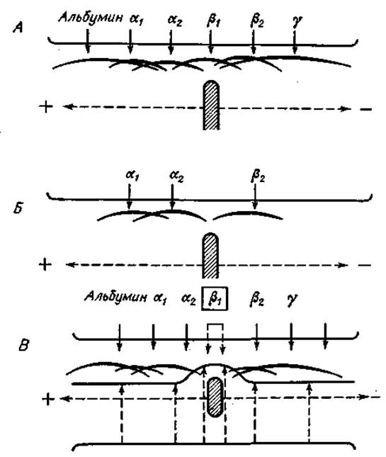
Fig. 31. Methods for analyzing protein mixtures by Immunoelectrophoresis (see text for details).
Scope of application. This method can be used to identify individual components of protein mixtures of unknown composition, provided that the researcher has the appropriate immune serum and a mixture of control (absorbing) antigens.
PROCEDURE
1. Preparation of the agar plate. Pour 40 ml of molten 1.5% agar in veronal buffer, pH 8.2, onto a 10 x 16 cm Glass plate. Cut the solidified agar gel layer longitudinally so that, after removing the excess gel, only a 15 mm wide strip remains on the glass. On the cathode side, at a distance of one-third of the length of this gel strip and equidistant from its edges, make a sample well approximately 5 mm in diameter and seal it with 1—2 drops of molten agar.
2. Electrophoresis. After loading the test sample, perform electrophoresis for 7—8 h at a voltage gradient of 3 V/cm.
3. Application of gel strips containing absorbing antigens and immune serum. Upon completion of electrophoresis, pour molten agar mixed with a solution of control (absorbing) antigens adjacent to the gel strip containing the separated antigens, to form a second strip 5 mm wide. After the applied agar solidifies, cast a third strip of agar gel containing the immune serum next to the second one. As a result, three adjacent strips of agar gel are formed on a single glass plate, representing: a) the specific immune serum zone, b) the absorbing antigen zone, and c) the zone of electrophoretically separated test antigens.
4. Recording of results. Precipitin bands develop over several days at 4°C in a humid chamber. Their appearance can be recorded by photographing native or stained preparations (see p. 135).
NOTES
1. This method is highly useful, for example, in diagnosing deficiencies in the synthesis of a particular class of IMMUNOGLOBULINS. In this case, normal human serum is subjected to electrophoretic separation, the patient's serum is introduced into the absorbing antigen zone, and antiserum to human Serum Proteins is placed in the immune serum zone. Only those antiglobulin antibodies that do not encounter their corresponding immunoglobulins in the patient's serum while diffusing from the antiserum zone to the normal serum zone participate in the formation of precipitin bands with the normal human serum fractions. Consequently, precipitin bands appear that correspond to the missing class of immunoglobulins, such as IgG, IgA, or IgM.
2. In the gel strip containing the absorbing antigens, straight precipitin bands appear at the sites of antigen-antibody complex formation.
Last update: 06/08/2026
Editorial and Educational Adaptation: This material has been compiled based on the primary/original source text. The project team performed an editorial review, corrected technical inaccuracies, structured sections, and adapted the content for an educational format.
What was processed:
- elimination of formatting defects (OCR errors, structural breaks, corrupted characters);
- editorial organization of content;
- standardization of terminology in accordance with academic sources;
- verification of factual statements against the original source text.
All mentions of the author, publication year, and origin of the primary text have been preserved in accordance with the source.