Review of Medical Physiology - William F. Ganong 2002
Endocrine System, Metabolism, and Reproduction
Adrenal Medulla and Adrenal Cortex
Adrenal Cortex - Transport, Metabolism, and Excretion of Adrenocortical Hormones
Glucocorticoid binding
In the bloodstream, cortisol is bound to an α-globulin called transcortin, or corticosteroid-binding globulin (CBG). It also partially binds to albumin (see Table 23-5). Corticosterone is bound in a similar manner, though to a lesser extent. Consequently, the half-life of cortisol in the bloodstream is longer (approximately 60-90 min) than that of corticosterone (50 min). Bound Steroids are physiologically inactive. Protein binding results in relatively small amounts of free cortisol and corticosterone in the urine.
The equilibrium between cortisol and its binding protein, as well as tissue cortisol and ACTH secretion, is shown in Fig. 20-12. The function of bound cortisol is to store the hormone in the Blood, ensuring a steady supply of free cortisol to Tissues. This relationship is similar to that of T4 and its binding protein (see Fig. 18-8). At a normal total cortisol level (13.5 μg/dL, or 375 nmol/L) in plasma, there is very little free cortisol, but CBG binding sites become fully saturated when total plasma cortisol exceeds 20 μg/dL. At higher concentrations, a slight increase in albumin binding is observed, but the main excess remains in the unbound form.
Class="center">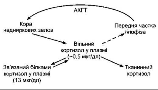
Fig. 20-12. Relationship between free and bound cortisol. The dashed arrow indicates that cortisol inhibits ACTH secretion. The free cortisol content is an approximate value from many measurements, calculated as the difference between total plasma cortisol and protein-bound cortisol.
CBG is synthesized in the Liver, and its production is enhanced by estrogens. CBG concentration increases during Pregnancy and decreases in cirrhosis, nephrosis, and myeloma. When CBG concentration rises, most of the cortisol becomes bound, initially decreasing the level of free cortisol. This stimulates ACTH secretion, leading to increased cortisol synthesis until a new equilibrium is reached, where The amount of bound cortisol is elevated but free cortisol is normal. The opposite changes occur when CBG concentration decreases. This explains why pregnant women have high total plasma cortisol concentrations without signs of glucocorticoid excess, and conversely, why some patients with nephrosis have low total plasma cortisol without signs of glucocorticoid deficiency.
METABOLISM and excretion of glucocorticoids
Cortisol metabolism occurs in the liver, which is the primary site of glucocorticoid Catabolism. Most cortisol is reduced to dihydrocortisol and then to tetrahydrocortisol, which is conjugated with glucuronic acid (Fig. 20-13). The glucuronyl transferase system responsible for this conversion also catalyzes The formation of bilirubin glucuronides (see Chapter 26), as well as many Hormones and drugs. There is competitive inhibition between these substrates for this enzyme system.
The Liver and other tissues contain the enzyme 11β-hydroxysteroid dehydrogenase (see below). There are at least two isoforms of this enzyme. Type 1 catalyzes the interconversion of cortisol and cortisone, although it Functions primarily as a reductase, forming cortisol from cortisone. Type 2 catalyzes almost exclusively the irreversible conversion of cortisol to cortisone.
Cortisone is an active glucocorticoid, well-known for its widespread medical use, but it is not synthesized in significant amounts by the Adrenal Glands. Only a small amount of the cortisone formed in the liver enters the Circulation because it is rapidly reduced and conjugated to form tetrahydrocortisone glucuronide. The tetrahydroglucuronide derivatives (conjugates) of cortisol and corticosterone are highly soluble. They enter the circulation, where they do not bind to Proteins, and are rapidly excreted in the urine, partly via tubular secretion.
About 10% of secreted cortisol is converted in the liver to 17-Ketosteroid derivatives of cortisol and cortisone. These ketosteroids are conjugated mainly with sulfate and then excreted in the urine. Other metabolites, including 20-hydroxy derivatives, are also formed.
Enterohepatic circulation of glucocorticoids also occurs, and about 15% of the secreted cortisol is excreted in the feces. Cortisone is metabolized similarly to cortisol, but it does not form 17-ketosteroid derivatives.
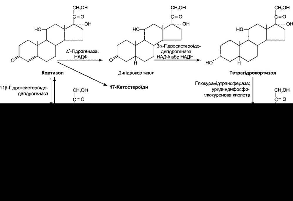
Fig. 20-13. Outline of cortisol metabolism in the liver.
Changes in the rate of hepatic metabolism
The rate of hepatic inactivation of glucocorticoids is reduced in liver disease and, interestingly, during surgery and other stresses. Consequently, in stressed individuals, the plasma concentration of free cortisol is higher than during maximal ACTH stimulation without stress.
Aldosterone
Aldosterone is only minimally bound to proteins, and its half-life is short (about 20 min). The amounts secreted are small (see Table 20-1), and the normal total plasma aldosterone concentration in humans is approximately 0.006 μg/dL (0.17 nmol/L). For comparison, the level of cortisol (bound and free) is approximately 13.5 μg/dL (375 nmol/L). Most aldosterone is converted in the liver to a tetrahydroglucuronide derivative, but some is converted to aldosterone 18-glucuronide. Unlike the metabolites of Other Steroids, this derivative is converted back to free aldosterone by Hydrolysis at pH = 1.0; hence, it is often referred to as the acid-labile conjugate. Less than 1% of secreted aldosterone is found in the urine in free form, 5% as the acid-labile conjugate, and over 40% as tetrahydroglucuronide.
17-Ketosteroids
The major adrenal androgen is the 17-ketosteroid dehydroepiandrosterone, although androstenedione is also formed. Both 11-hydroxy derivatives of androstenedione and 17-ketosteroids are formed from cortisol and cortisone by side-chain Cleavage in the liver. They are the only 17-ketosteroids that possess an =O or -OH group at position 11 (11-oxy-17-ketosteroids). Testosterone is also metabolized to 17-ketosteroids. Since the daily excretion of 17-ketosteroids in adults is 15 mg for men and 10 mg for women, about two-thirds of urinary ketosteroids in humans are secreted by the adrenal glands or formed from cortisol in the liver, while about one-third are of testicular origin.
Etiocholanolone, one of the metabolites of adrenal androgens and testosterone, can cause fever when unconjugated (see Chapter 14). Some individuals experience episodic fevers due to the periodic accumulation of unconjugated etiocholanolone in the blood (etiocholanolone fever).
Last update: 10/08/2026
Editorial and Educational Adaptation: This material has been compiled based on the primary/original source text. The project team performed an editorial review, corrected technical inaccuracies, structured sections, and adapted the content for an educational format.
What was processed:
- elimination of formatting defects (OCR errors, structural breaks, corrupted characters);
- editorial organization of content;
- standardization of terminology in accordance with academic sources;
- verification of factual statements against the original source text.
All mentions of the author, publication year, and origin of the primary text have been preserved in accordance with the source.