Orthopedics - Oleksa A.P. 2006
Examination of the orthopedic patient
Measurement of limb length
Very often, when examining patients, it is necessary to determine the length of the limbs and their individual segments. This measurement is performed using a tape measure by comparing the affected limb with the healthy one. It reflects the degree of limb shortening or lengthening in centimeters.
Measurements are taken with the patient lying on a firm couch. The limbs are positioned symmetrically with correct alignment of the trunk: the line connecting the acromia and the line connecting the anterior superior iliac spines must be perpendicular to the midline of the body.
The length of the upper limb is defined as the distance from the acromion of the scapula to the tip of the distal phalanx of the third digit of the straightened hand (Fig. 11). The anatomical (true) length of a limb is measured segment by segment, so its exact posture is not critical. However, to better palpate bony landmarks, it is advisable to keep the limb semi-flexed.
Limb shortening can be caused by bone fractures with angular or rotational fragment displacement, bone dislocations, inflammatory, degenerative-dystrophic, and destructive joint processes, as well as flexion-adduction contractures and ankylosis in a malposition.
An increase in limb length occurs during growth As a result of irritation of the epiphyseal growth plate (due to an inflammatory bone process, a foreign body, metal hardware, etc.), as well as from an abnormal limb posture (such as an abduction contracture of the hip).
The following types of limb shortening or lengthening are distinguished:
1. True, or anatomical, shortening (lengthening) is determined by measuring the absolute length of a specific anatomical bone segment of the affected limb compared to the healthy one. The true length of the thigh is measured (Fig. 12) from the tip of the greater trochanter to the outer joint line of the knee; that of the leg, from the medial joint line of the knee joint to the tip of the medial malleolus; that of the arm, from the acromion or greater tubercle of the humerus to its lateral epicondyle; and that of the forearm, from the tip of the olecranon process of the ulna to its styloid process.
Class="center">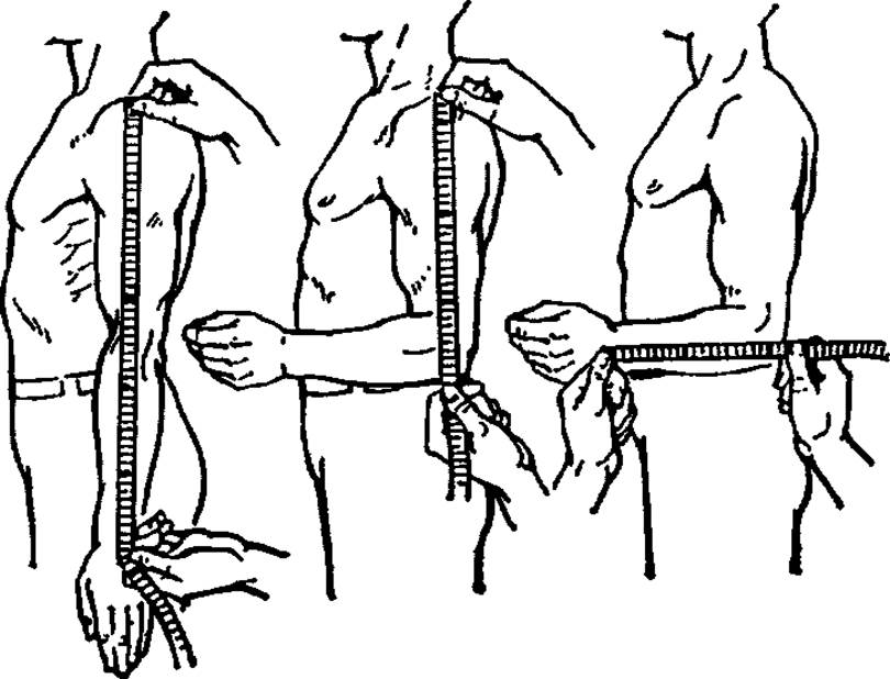
Fig. 11. Measurement of upper limb length.
2. Positional, or apparent, shortening (lengthening) of a limb (Fig. 13) develops as a result of joint contracture or ankylosis with malposition of the adjacent segment. For example, a flexion-adduction contracture of the hip or ankylosis of the hip joint in a similar position leads to lower limb shortening, even though the anatomical length of its individual segments is normal upon measurement. Such shortening is termed positional because the levels of the knee joints and feet of the healthy and shortened limbs do not coincide projectively.
3. Relative, or dislocation-related, shortening (lengthening) of a limb (Fig. 14) in superior-posterior dislocation is caused by the displacement of one of its segments. For example, the Displacement of the femoral HEAD upward, beyond the Roser-Nelaton line, leads to limb shortening, although the anatomical lengths of the Femur and Tibia remain equal.
4. Total, or clinical (functional), shortening (lengthening) of a limb (Fig. 15) is caused by the combination of the aforementioned types of shortening or lengthening. For example, adult patients with congenital hip dislocation may simultaneously exhibit all types of limb shortening: relative, due to the displacement of the femoral head outside the acetabulum; positional, resulting from flexion-adduction contracture; and anatomical, affecting individual segments due to stunted growth of the abnormally functioning limb bones. Total shortening of the lower limb should be measured with the patient standing, by placing wooden blocks under the FOOT of the shortened leg until pelvic obliquity is corrected—that is, until its upper spines are parallel to the floor. For this purpose, it is convenient to have a set of supporting blocks with thicknesses of 0.5, 1, 2, 3, and 5 cm.
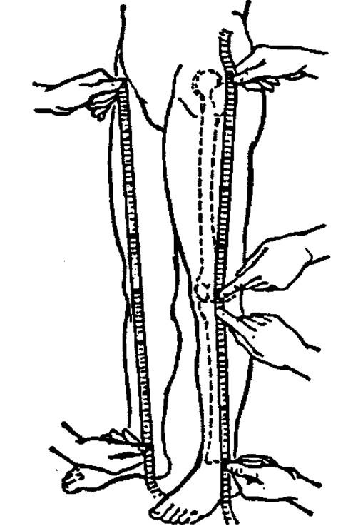
Fig. 12. Measurement of lower limb length.
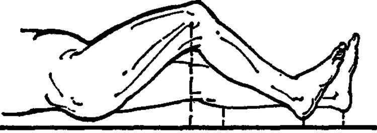
Fig. 13. Apparent shortening of the lower limb.
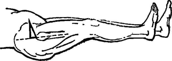
Fig. 14. Relative (dislocation) shortening of the lower limb.
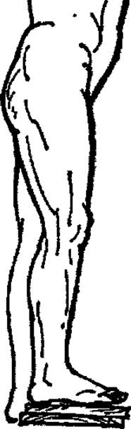
Fig. 15. Measurement of total (functional) shortening of the lower limb.
Last update: 10/08/2026
Editorial and Educational Adaptation: This material has been compiled based on the primary/original source text. The project team performed an editorial review, corrected technical inaccuracies, structured sections, and adapted the content for an educational format.
What was processed:
- elimination of formatting defects (OCR errors, structural breaks, corrupted characters);
- editorial organization of content;
- standardization of terminology in accordance with academic sources;
- verification of factual statements against the original source text.
All mentions of the author, publication year, and origin of the primary text have been preserved in accordance with the source.