Human Anatomy Part 2 - K. A. Dyubenko, A. K. Kolomiytsev, Yu. B. Chaykovsky 2008
Special Section
Sense organs, organa sensuum
Vestibulocochlear organ - External ear
The Vestibulocochlear Organ, organum vestibulocochleare, consists of the external, middle, and internal ear (auris externa, media et interna)
The External ear comprises the auricle, auricula, and the external acoustic meatus, meatus acusticus externus (Fig. 220). The framework of the auricle is formed by elastic Cartilage, cartilago auricularis, covered with thin Skin. The latter adheres tightly to the cartilage, meaning subcutaneous fat is absent here. The lower part of the auricle is the lobule, lobulus auricularis, which lacks a cartilaginous framework and is a skin pouch containing adipose tissue.
The auricle, together with the acoustic meatus, acts as an acoustic funnel for catching sounds. The convex surface of the auricle faces medially and backwards, while the concave surface faces laterally and forwards. The free margin of the auricle is rolled, forming the helix. The anterior end of the helix is termed the crus of the helix, crus helicis, and the posterior end is the tail, cauda helicis. Running parallel to the helix is an elevation called the antihelix, antihelix, which superiorly divides into two crura, crura antihelicis. They enclose the triangular fossa, fossa triangularis. The helix and antihelix are separated from each other by the scapha, scapha. Anterior to the antihelix lies a depression—the concha of the auricle, concha auricularis, which is divided by the crus of the helix into an upper part, the cymba conchae, cymba conchalis, and a lower part, the cavum conchae, cavitas conchalis. At the bottom of the latter lies the external acoustic opening, poms acusticus extemus. Anteriorly, the conchal cavity is bounded by a projection, the tragus, tragus. Posteriorly, at the lower end of the antihelix, opposite the tragus, lies a tubercle—the antitragus, antitragus. The tragus and antitragus are separated by the intertragic notch, incisura intertragica. In the superior-posterior region of the auricle, a small tubercle (Darwin's) can sometimes be found, tuberculum auriculare. The cartilage of the auricle is attached to the Temporal bone by three ligaments—the anterior, superior, and posterior, ligg. auricularia anterius, superius, posterius. The auricular Muscles also attach to the cartilage of the auricle, although the latter do not play a significant functional role in humans.
The external acoustic meatus is located between the auricle and the tympanic membrane. Its lateral part consists of cartilage, cartilago meatus acustici, whereas the medial part has bony walls. The cartilaginous part curves forward and downward; therefore, during otoscopy, the physician straightens it by pulling the auricle backward and upward.
The junction area between the cartilaginous and bony parts is the narrowest section of the external acoustic meatus. On the anterior and inferior walls here, there are 2–5 clefts closed by Connective Tissue. Through these clefts, pus from the parotid gland can penetrate into the external acoustic meatus during purulent parotitis.
Class="center">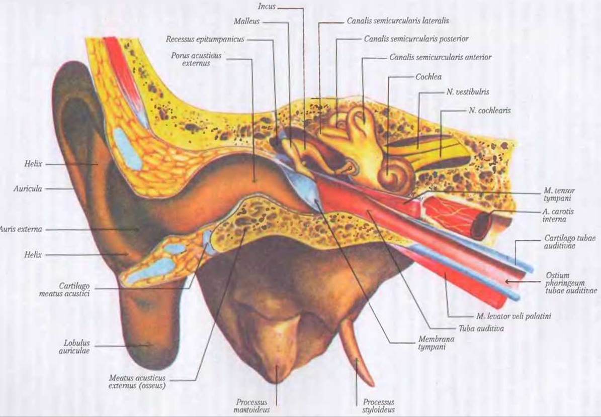
Fig. 220. Vestibulocochlear organ, organum vestibulocochleare (external, middle, and internal ear)
The external acoustic meatus is lined internally with thin perichondrium and periosteum, and covered externally with skin. In the region of the cartilaginous part, the skin contains numerous coarse auricular hairs, Sebaceous Glands, and specialized tubular glands—modified Sweat Glands known as ceruminous glands, glandulae cerwninosae, which secrete earwax, cerumen.
The Blood supply to the external ear is well developed and provided by the occipital, posterior auricular, and superficial temporal Arteries, a. temporalis superficialis. However, the absence of adipose tissue around the arteries makes the auricle highly sensitive to low ambient temperatures. The bony part of the acoustic meatus is supplied by the deep auricular artery, a. auricularis profunda. Veins correspond to the arteries. Lymph drains into the parotid and deep cervical Lymph Nodes, nodi limphatici parotidei et cervicales profundi.
The Innervation of the external ear is complex. The anterior part of the auricle, along with the anterior and superior walls of the external acoustic meatus, is innervated by the auriculotemporal nerve, n. auriculo-temporalis; the posterior surface of the auricle by the great auricular and lesser occipital nerves, n. auricularis magnus et n. occipitalis minor; and the concave part of the auricle, as well as the posterior and inferior walls of the external acoustic meatus, by the vagus, glossopharyngeal, and intermediofacial nerves, nervus vagus, n. glossopharyngeus et n. intermediofacialis.
Last update: 08/08/2026
Editorial and Educational Adaptation: This material has been compiled based on the primary/original source text. The project team performed an editorial review, corrected technical inaccuracies, structured sections, and adapted the content for an educational format.
What was processed:
- elimination of formatting defects (OCR errors, structural breaks, corrupted characters);
- editorial organization of content;
- standardization of terminology in accordance with academic sources;
- verification of factual statements against the original source text.
All mentions of the author, publication year, and origin of the primary text have been preserved in accordance with the source.