MEDICAL BIOLOGY, ANATOMY, HUMAN PHYSIOLOGY AND PATHOLOGY - Y.I. Fedoniuk 2010
ANATOMY, PHYSIOLOGY, PATHOLOGY
CHAPTER 9. REPRODUCTIVE PROCESS
ANATOMY OF THE REPRODUCTIVE ORGANS
MALE REPRODUCTIVE ORGANS
8. PENIS. MALE URETHRA
The Penis consists of two corpora cavernosa and one corpus spongiosum. The corpus spongiosum lies ventral to the corpora cavernosa and is traversed by the Urethra. The posterior part of the penis is called the ROOT, and the anterior, expanded part is the glans. The middle portion forms the body (shaft) of the penis. The external urethral orifice is located on the glans. The Skin near the glans forms a fold known as the prepuce (foreskin). Its inner surface contains Sebaceous Glands, the secretion of which is called smegma. The outer envelope of the corpora cavernosa is a dense fibrous tunica albuinea, from which numerous trabeculae extend inward. The spaces between the trabeculae are filled with Blood. The size of the penis varies depending on the volume of blood within the cavernous bodies.
The male urethra (urethra masculina) is an unpaired tubular organ approximately 16–22 cm long with a diameter of 0.5–0.7 cm (Fig. 9.3). It serves for The excretion of urine and semen. The urethra extends from the Urinary Bladder to the external orifice on the glans penis. It is divided into three parts: the prostatic part (passing through the Prostate Gland), the membranous part (passing through the urogenital Diaphragm), and the spongy ( penile) part. Surrounded by the corpus spongiosum, the prostatic part is the widest, whereas the membranous part is the narrowest. The urethra has an S-shaped curve. When a catheter is introduced into the urethra, its anterior curvature is straightened, leaving only the posterior, more fixed curvature. The mucous membrane of the urethra is lined in different regions by various types of epithelium: transitional, pseudostratified columnar, simple columnar, and stratified squamous. The muscular layer consists of circular and longitudinal smooth Muscle fibers.
Class="center">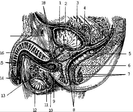
Fig. 9.3. Median sagittal section of the male pelvis
Last update: 08/08/2026
Editorial and Educational Adaptation: This material has been compiled based on the primary/original source text. The project team performed an editorial review, corrected technical inaccuracies, structured sections, and adapted the content for an educational format.
What was processed:
- elimination of formatting defects (OCR errors, structural breaks, corrupted characters);
- editorial organization of content;
- standardization of terminology in accordance with academic sources;
- verification of factual statements against the original source text.
All mentions of the author, publication year, and origin of the primary text have been preserved in accordance with the source.