MEDICAL BIOLOGY, HUMAN ANATOMY, PHYSIOLOGY AND PATHOLOGY - Y.I. Fedoniuk 2010
ANATOMY, PHYSIOLOGY, PATHOLOGY
SECTION 6. THE RESPIRATORY PROCESS AND ITS PATHology
PLEURA
The Pleura is a serous membrane that covers the Lungs on all sides and, in the region of the lung ROOT, reflects onto the walls of the thoracic cavity, forming a closed pleural sac around each lung (Fig. 6.9).
Class="center">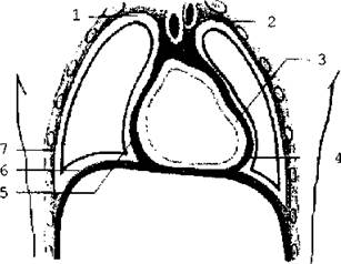
Fig. 6.9. Diagram of the pleural and pericardial cavities (frontal section).
The layer of pleura that lines the walls of the thoracic cavity is called the parietal pleura. Depending on the area it covers, the costal, diaphragmatic, and mediastinal pleura are distinguished.
The pleura that covers the lungs and fuses with their surface is called the visceral (pulmonary) pleura. Between these two layers lies the pleural cavity, which contains 1-2 ml of serous fluid to reduce friction between the membranes during Respiration. In areas where one part of the parietal pleura transitions into another, pleural recesses (sinuses) are formed: the costomediastinal, costodiaphragmatic, and phrenicomediastinal sinuses.
These spaces are filled by the lungs during deep inspiration. At rest, their walls lie closely apposed to one another. The costodiaphragmatic recess is the largest of them.
Last update: 08/08/2026
Editorial and Educational Adaptation: This material has been compiled based on the primary/original source text. The project team performed an editorial review, corrected technical inaccuracies, structured sections, and adapted the content for an educational format.
What was processed:
- elimination of formatting defects (OCR errors, structural breaks, corrupted characters);
- editorial organization of content;
- standardization of terminology in accordance with academic sources;
- verification of factual statements against the original source text.
All mentions of the author, publication year, and origin of the primary text have been preserved in accordance with the source.