MEDICAL BIOLOGY, HUMAN ANATOMY, PHYSIOLOGY AND PATHOLOGY - Ya.I. Fedoniuk 2010
ANATOMY, PHYSIOLOGY, PATHOLOGY
CHAPTER 2. MUSCULOSKELETAL SYSTEM (THE MECHANISM OF MOVEMENT)
MUSCLES AND FASCIAE OF BODY REGIONS
MUSCLES AND FASCIAE OF THE UPPER LIMB
2. MUSCLES OF THE FREE UPPER LIMB
The Muscles of the arm are divided into anterior (flexors) and posterior (extensors) groups (Fig. 2.90).
Class="center">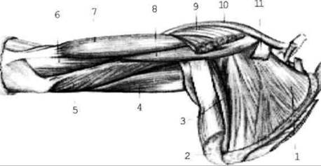
Fig. 2.90. Muscles of the shoulder girdle and arm; anterior view, the pectoralis Major and minor muscles have been removed
The anterior group includes the biceps brachii, brachialis, and coracobrachialis muscles.
1. The biceps brachii Muscle (biceps), m. biceps brachii, originates by two heads: the long HEAD from the supraglenoid tubercle of the scapula and the short head from the coracoid process of the scapula. It inserts into the radial tuberosity. Function: flexes the arm and forearm.
2. The brachialis muscle (m. brachialis) originates from the humerus and inserts into the ulnar tuberosity. Function: flexes the forearm.
3. The coracobrachialis muscle (m. coracobrachialis) extends from the coracoid process of the scapula and inserts into the humerus. Function: flexes and adducts the arm.
The posterior group includes the triceps brachii and anconeus muscles.
The triceps brachii muscle (m. triceps brachii) has three heads: medial, lateral (which originate from the humerus), and long (originating from the infraglenoid tubercle of the scapula). It inserts via a common tendon into the olecranon of the ulna. Function: extends the forearm.
The anconeus muscle is small and may occasionally be absent. Function: assists the preceding muscle in extending the forearm.
The muscles of the forearm are divided into anterior (flexors) and posterior (extensors) groups. The muscles of the anterior group are further subdivided into superficial and deep layers. The superficial muscles originate mainly from the medial epicondyle of the humerus. The deep muscles originate from various points on the radius and ulna.
Functionally, all muscles of the anterior group act as flexors of the wrist and fingers, as well as pronators.
The anterior group includes:
1) pronator teres;
2) flexor carpi radialis;
3) flexor carpi ulnaris;
4) flexor digitorum superficialis;
5) palmaris longus;
6) brachioradialis.
Deep layer (Fig. 2.91 b):
7) deep flexor of the fingers;
8) pronator quadratus;
9) long flexor of the thumb
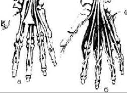
Fig. 2.91. Superficial (a) and deep (b) muscle layers
Muscles of the posterior compartment
originating from the lateral epicondyle of the humerus and bony landmarks of the radius and ulna, they are divided into extensors of the wrist and fingers, as well as supinators.
Superficial muscles (Fig. 2.92): 1) extensor carpi radialis longus and brevis; 2) extensor digitorum; 3) extensor carpi ulnaris.
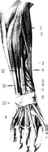
Fig. 2.92. Muscles of the posterior aspect of the forearm, right upper extremity
Deep muscles: 1) supinator; 2) abductor pollicis longus; 3) extensor pollicis brevis and longus; 4) extensor indicis.
The Muscles of the hand are located exclusively on the palmar surface. They are divided into three groups: thenar muscles, hypothenar muscles, and the central palmar group (Fig. 2.93).
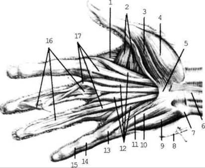
Fig. 2.93. Muscles of the right hand; palmar surface (fascia partially removed):
Thenar muscles: 1) flexor pollicis brevis; 2) abductor pollicis brevis; 3) opponens pollicis (opposes the thumb to all other fingers); 4) adductor pollicis.
Hypothenar muscles: 1) abductor digiti minimi; 2) flexor digiti minimi brevis, and 3) opponens digiti minimi.
The central group (of the palmar depression) comprises four lumbricals (which flex the proximal Phalanges and extend the middle and distal ones) and the interossei. The latter fill the metacarpal spaces and are divided into PALMAR AND DORSAL. The palmar interossei adduct all fingers toward the middle finger, while the dorsal interossei abduct the fingers.
Last update: 08/08/2026
Editorial and Educational Adaptation: This material has been compiled based on the primary/original source text. The project team performed an editorial review, corrected technical inaccuracies, structured sections, and adapted the content for an educational format.
What was processed:
- elimination of formatting defects (OCR errors, structural breaks, corrupted characters);
- editorial organization of content;
- standardization of terminology in accordance with academic sources;
- verification of factual statements against the original source text.
All mentions of the author, publication year, and origin of the primary text have been preserved in accordance with the source.