WORKSHOP IN HISTOLOGY, CYTOLOGY AND EMBRYOLOGY - 2016
Chapter 5. SPECIAL HISTOLOGY
Nervous system and sense organs.
Slide 67. Sensory (spinal) ganglion.
Staining: hematoxylin and eosin.
Magnification: x400.
The slide represents a Cytology/practical/54.html">Longitudinal section of the ganglion.
At low magnification, we can see the ganglion capsule (1) formed by Connective Tissue. Within the ganglion, we distinguish Neurons (2) and nerve fibers (3). The perikarya of peripheral pseudounipolar neurons are located mainly beneath the capsule, while the fibers occupy the central part of the ganglion. The neurons are rounded in shape with a large vesicular Nucleus, surrounded by mantle gliocytes (4), which can be identified by their round nuclei surrounding the neurons. Layers of delicate Fibrous connective tissue are visible externally.
The slide also shows the dorsal (5) and ventral (6) roots of the Spinal Cord.
Class="center">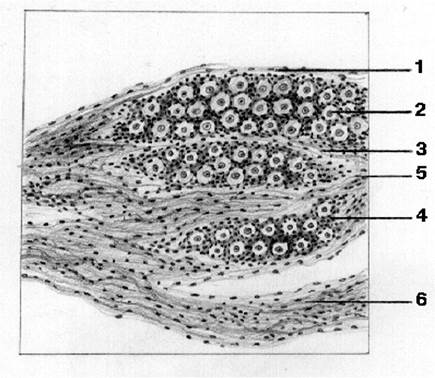
Slide 67. Sensory (spinal) ganglion.
Last update: 07/08/2026
Editorial and Educational Adaptation: This material has been compiled based on the primary/original source text. The project team performed an editorial review, corrected technical inaccuracies, structured sections, and adapted the content for an educational format.
What was processed:
- elimination of formatting defects (OCR errors, structural breaks, corrupted characters);
- editorial organization of content;
- standardization of terminology in accordance with academic sources;
- verification of factual statements against the original source text.
All mentions of the author, publication year, and origin of the primary text have been preserved in accordance with the source.