WORKSHOP IN HISTOLOGY, CYTOLOGY AND EMBRYOLOGY - 2016
Chapter 4. TISSUES
Slide 36. Chick embryo mesenchyme.
Staining: hematoxylin and eosin.
Magnification: x400.
Mesenchyme is the most primitive Connective Tissue during Cytology/cytology/16.html">Early stages of embryonic development. It is formed by stellate or spindle-shaped Cells (1) with round or oval nuclei. The cells contact each other via their processes, forming a network (2). The intercellular amorphous substance (3) is transparent, unstained, and fiber-free. Occasionally, clusters of mesenchymal cells can be observed, which subsequently give rise to Blood Vessels.
Class="center">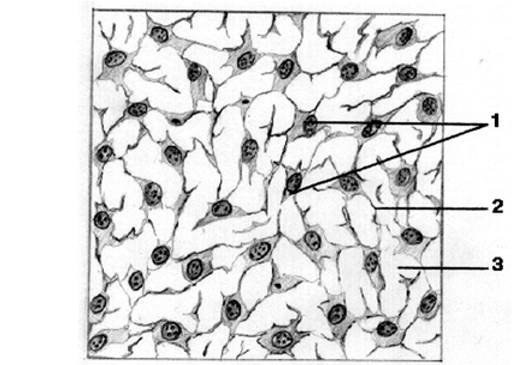
Slide 36. Chick embryo mesenchyme.
Last update: 07/08/2026
Editorial and Educational Adaptation: This material has been compiled based on the primary/original source text. The project team performed an editorial review, corrected technical inaccuracies, structured sections, and adapted the content for an educational format.
What was processed:
- elimination of formatting defects (OCR errors, structural breaks, corrupted characters);
- editorial organization of content;
- standardization of terminology in accordance with academic sources;
- verification of factual statements against the original source text.
All mentions of the author, publication year, and origin of the primary text have been preserved in accordance with the source.