BIOLOGY Volume 2 - A Guide to General Biology - 2004
14. TRANSPORT IN ANIMALS
14.7. The Heart
14.7.2. The Cardiac Cycle
The cardiac cycle is The sequence of events occurring during one heartbeat. It includes the contraction (systole) and relaxation (diastole) of The Heart Muscle. The cycle consists of the following phases.
1. Atrial diastole. Both the atria and ventricles are relaxed, and Blood flows into the atria from the Veins under low pressure: oxygenated (arterial) blood into the left atrium, and deoxygenated (venous) blood into the right atrium. Initially, the bicuspid and tricuspid Valves are closed (Fig. 14.17, A), but as the atria fill with blood, the pressure within them rises (Fig. 14.18). Eventually, it exceeds the pressure in the relaxed ventricles, forcing these valves to open.
2. Atrial systole. As diastole ends, both atria contract simultaneously, pumping blood into the ventricles (Fig. 14.17, B).
3. Ventricular systole. Almost simultaneously with the atria (0.1–0.2 s later), the ventricles contract (Fig. 14.17, C). This increases intraventricular pressure, closing the atrioventricular valves to prevent backflow into the atria. At the same time, it opens the semilunar valves, forcing blood into the pulmonary artery and the aorta. The sharp snapping shut of the atrioventricular valves during ventricular systole produces a distinct sound (approximately "lub"); this is known as the first (systolic) heart sound.
4. Ventricular diastole. Ventricular systole is followed by ventricular diastole (Fig. 14.17, D). Due to the high pressure generated in the aorta and pulmonary artery, some blood surges back toward the ventricles. Blood fills the semilunar pockets, causing the valves to snap shut, thereby preventing blood from returning to the ventricles. The sharp closure of the semilunar valves produces a sound resembling "dub". This is the second (diastolic) heart sound.
Class="center">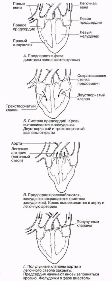
Fig. 14.17. Sequence of phases in a single cardiac cycle.

Fig. 14.18. Sequence of events throughout the cardiac cycle, starting with atrial systole. A portion of the next cycle is also shown. PA — blood pressure in the pulmonary artery; RA — in the right atrium; LA — in the left atrium; ECG — Electrocardiogram; P, Q, R, S, T — standard designations for electrocardiographic waves. (From J. H. Green (1968). An Introduction to human physiology, Oxford University Press.).
During ventricular systole, the walls of elastic Arteries stretch, and during diastole, they recoil to their original state and propel the blood forward. As a result, Blood flow through the SYSTEMIC AND PULMONARY circulations is pulsatile. As blood moves further from the heart, the pulse gradually weakens until it disappears entirely in the capillaries and veins, where blood flow becomes steady (Figs. 14.19 and 14.20). Thus, one complete cycle consists of one systole and one diastole and lasts approximately 0.8 s (Fig. 14.18).

Fig. 14.19. Distribution of blood volume, pressure, and flow velocity in the human Circulatory system. (From K. Schmidt-Nielsen (1980). Animal physiology, 2nd ed., Cambridge University Press.).

Fig. 14.20. Blood pressure in various sections of the human circulatory system. (J. H. Green (1968). An introduction to human physiology, Oxford University Press.)
Last update: 06/08/2026
Editorial and Educational Adaptation: This material has been compiled based on the primary/original source text. The project team performed an editorial review, corrected technical inaccuracies, structured sections, and adapted the content for an educational format.
What was processed:
- elimination of formatting defects (OCR errors, structural breaks, corrupted characters);
- editorial organization of content;
- standardization of terminology in accordance with academic sources;
- verification of factual statements against the original source text.
All mentions of the author, publication year, and origin of the primary text have been preserved in accordance with the source.