Practical Protein Chemistry - A. Darbre 1989
X-ray Crystallography and Electron Microscopy
Electron Microscopy
Three-Dimensional Electron Microscopy
The images we observe under a Microscope are two-dimensional projections of the three-dimensional Structure OF THE specimen under investigation. By using a goniometer—a device that tilts the specimen within the electron beam—it can be examined from various angles. A series of such images provides significantly more structural information than any single image alone. The most informative approach, however, is to reconstruct the three-dimensional structure of the object based on these images. For certain specimens, this is indeed feasible, although the process is quite time-consuming. The mathematical framework employed here is analogous to that of X-ray crystallography. The micrographs of the studied object, taken at different angles, are converted into numerical form using a densitometer and entered into computer memory. The computer-generated three-dimensional image of the structure can be visually displayed in various ways. Typically, it is presented as a series of sections, similar to those obtained with a microtome for histological samples embedded in a polymer matrix [3, 4].
To date, high-quality three-phase electron density maps have been obtained for several well-ordered "stained" objects. Fig. 20.17 illustrates such a map calculated for ribosome crystals [14]. Such maps are analogous to low-resolution X-Ray Diffraction maps (5 nm).
Class="center">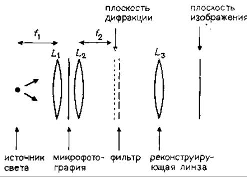
FIG. 20.15. An optical diffractometer in which a photomicrograph is illuminated by a parallel beam of light, thereby generating a diffraction pattern. A filter and a lens positioned downstream of the diffraction pattern reconstruct the filtered image.
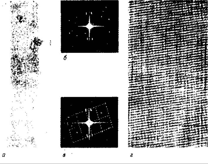
FIG. 20.16. Optical filtering of a two-layered structure image: a — photomicrograph of an Actin tube; b — diffraction pattern of the photomicrograph containing maxima from both layers and representing a superposition of two reciprocal lattices; c — lattice of diffraction maxima selected for reconstruction and indicated by dashed lines; d — filtered image reconstructed from the selected lattice.
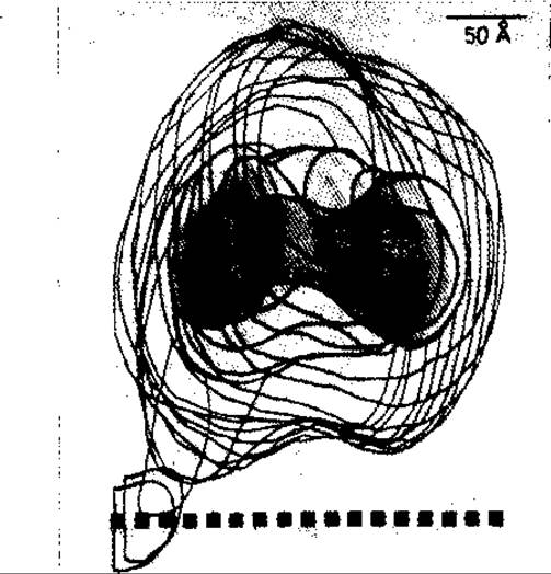
FIG. 20.17. Three-dimensional reconstruction of a ribosome crystal unit Cell based on a series of micrographs taken at different angles. The Ribosomes were found in the crystalline state within oocytes of the lizard Lacerta sicula. L and S denote the large and small ribosomal subunits, respectively.
Last update: 06/08/2026
Editorial and Educational Adaptation: This material has been compiled based on the primary/original source text. The project team performed an editorial review, corrected technical inaccuracies, structured sections, and adapted the content for an educational format.
What was processed:
- elimination of formatting defects (OCR errors, structural breaks, corrupted characters);
- editorial organization of content;
- standardization of terminology in accordance with academic sources;
- verification of factual statements against the original source text.
All mentions of the author, publication year, and origin of the primary text have been preserved in accordance with the source.