Practical Protein Chemistry - A. Darbre 1989
X-ray crystallography and electron microscopy
Molecular imaging as a method of structural analysis
When examining a specimen under a conventional Light Microscope, the light scattered by the object is focused by the objective lens to form a magnified image (Fig. 20.1). When light waves are scattered by the specimen, they undergo phase changes; as the objective lens focuses them, it preserves this post-scattering phase relationship. Consequently, the recombination of these rays forms an image of the object (Fig. 20.2). Resolution—that is, the minimum size of details reproducible in such an image—is limited by the wavelength of the light used (typically ∼600 nm). Finer details could theoretically be resolved using a microscope operating, for example, on X-rays with a wavelength of 0.154 nm (CuKa line). Unfortunately, lenses capable of focusing X-rays have not yet been developed, making direct image formation under such conditions impossible. One can only record the intensities of the rays scattered by the object under study, for instance, using a photographic plate. However, the phases of these rays remain unknown, making it impossible to reconstruct the image of the object.
Class="center">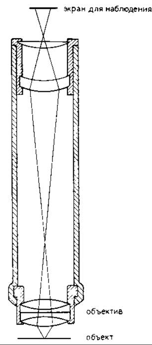
FIG. 20.1. Schematic diagram of a light microscope equipped with Glass lenses that collect the light scattered by the specimen and reconstruct a magnified image of the object. This design preserves the initial phase relationships of the scattered rays.
The object considered thus far could consist of many molecules in various orientations, such as protein molecules in solution. An image of such a specimen would depict each molecule in its specific orientation. However, by collecting data solely on ray intensities, it is impossible to isolate the rays scattered by a particular molecule from those scattered by the surrounding solvent molecules. This can be achieved by using crystalline samples, in which all molecules share the same orientation (Fig. 20.3). Due to the regularity of the crystal, the rays scattered by these molecules are concentrated into distinct diffraction peaks, making it possible to separate the radiation scattered by the protein molecules from Background scattering caused, for example, by solvent molecules, thereby significantly improving the signal-to-noise ratio. Investigating crystalline samples yields only an averaged image of the substance molecule within the crystal lattice, rather than an individual image of a specific molecule freely floating in solution. The diffraction pattern of a crystal (see, for example, Fig. 20.4) can be recorded using a suitably designed X-ray camera. The diffraction pattern reflects the three-dimensional Structure OF THE crystal under study. Therefore, a complete dataset of scattered ray intensities must include a series of such diffraction patterns stacked one above the other to form the so-called three-dimensional reciprocal lattice. The distances between the nodes of this lattice are inversely proportional to the intermolecular distances in the crystal. The intensities of the diffraction maxima, or spots, contain information about the Fine Structure of the molecule. If one could determine the relative phase values of the waves upon reaching the photographic plate, the molecular structure could be calculated, thereby yielding its image. However, determining the phases—or solving the so-called phase problem—remains one of the most challenging stages of X-ray crystallographic analysis.
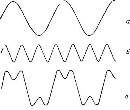
FIG. 20.2. Two light waves (a and b) with different amplitudes and phases, and their sum (c). It is evident that both amplitude and phase information are required for summation.
Last update: 06/08/2026
Editorial and Educational Adaptation: This material has been compiled based on the primary/original source text. The project team performed an editorial review, corrected technical inaccuracies, structured sections, and adapted the content for an educational format.
What was processed:
- elimination of formatting defects (OCR errors, structural breaks, corrupted characters);
- editorial organization of content;
- standardization of terminology in accordance with academic sources;
- verification of factual statements against the original source text.
All mentions of the author, publication year, and origin of the primary text have been preserved in accordance with the source.