Amino Acids, Peptides and Proteins - Devényi T., Gergely J. 1976
Protein analysis using low-voltage electrophoresis
Starch gel electrophoresis
Vertical starch gel electrophoresis
Principle of the method. Proteins migrate through a starch gel under METABOLISM/18.html">The Influence of an electric field.
Scope of application. Investigation of Serum proteins, lipo- and Glycoproteins, protein Hormones, Hemoglobins, tissue proteins, etc.
APPARATUS
1. Electrophoresis apparatus proposed by Smithies in 1955 [26]. Fig. 7 shows a schematic diagram of the apparatus for vertical starch gel electrophoresis. The vertically positioned tray containing the starch gel has dimensions of 6.5 x 20 x 250 mm. The buffer solution is poured into reservoirs 1 and 2, which are divided into two compartments by a Plexiglas partition. Carbon or platinum electrodes are located in the outer compartment of each reservoir. Both compartments in each reservoir, as well as the buffer solution and the starch gel, are connected to each other by wet strips of filter paper.
2. Power source: see p. 46.
Class="center">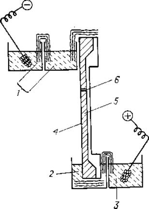
Fig. 7. Apparatus for starch gel electrophoresis (after Smithies [26]).
1, 2, 3 — reservoirs for buffer solution; 4 — plastic tray; 5 — starch gel; 6 — Sample application site.
PROCEDURE
1. Preparation of buffer solution. For the starch gel, a borate buffer solution of 0.03 M H3BO3 in 0.012 N NaOH is prepared, and for filling the Chambers of the apparatus, a buffer solution containing 0.3 M Н3ВО3 and 0.06 N NaOH is used.
2. Preparation of starch gel. A weighed portion of potato starch is suspended in 3 volumes of Water, mixed thoroughly, and allowed to settle for 30 min. The supernatant, containing soluble impurities from the starch, is discarded. Washing the starch by sedimentation is repeated twice, then the precipitate is transferred to a Büchner funnel and washed 3–4 times with buffer solution. After that, a paste-like mixture of starch and buffer solution is prepared, and approximately 100 ml of this paste is transferred to a 0.5 L Erlenmeyer flask. With constant stirring, the starch paste is heated until an absolutely homogeneous mass is obtained, avoiding boiling of the gel. To remove air bubbles, a vacuum is created in the flask containing the heated homogeneous starch gel using a vacuum pump, causing the gel to boil for 1–2 min. The gel is then used to fill the tray of the electrophoresis apparatus; excess buffer solution is removed with thick filter paper, and to prevent evaporation, The surface of the gel is covered with a thin layer of molten paraffin.
3. Connecting the internal electrical circuit of the apparatus. The starch gel in the vertically positioned tray and the buffer solution filling reservoirs 1 and 2, as well as the buffer and electrode compartments in each reservoir, are connected to each other by paper bridges. Each bridge consists of 10 strips of filter paper moistened with buffer solution.
4. Sample application. At a distance of 7 cm from the cathode end of the starch block, the sample application site is marked, and a 10 x 3 mm window is cut in the paraffin film. The layer of starch gel beneath the paraffin is then removed to form a slot into which the sample is introduced. This slot can be very conveniently made using the device shown in Fig. 8. It consists of a sharp-edged steel frame 1, which is pressed into the starch gel; the piece of gel that enters its inner cavity is then removed. 0.05 ml of serum is thoroughly mixed with dry starch in an amount sufficient to obtain a homogeneous paste of the same consistency as the gel itself. Using the plunger part 2 of the device, this paste is introduced into the slot. The frame should be removed very carefully to avoid damaging the integrity of the gel. Afterwards, the window in the paraffin film is sealed with a new layer of molten paraffin.
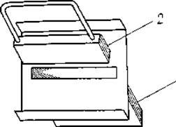
Fig. 8. Device used to make the slot for sample application into the starch block (see text for description).
5. Electrophoresis. Starch gel electrophoresis is carried out for 14–18 h at a voltage gradient of 6 V/cm.
6. Localization of protein fractions. After electrophoresis is completed, the starch gel tray is removed from the apparatus and placed horizontally on the bench. The paraffin film covering the gel is removed, and the entire starch surface is covered with a sheet of filter paper. The sheet is pressed firmly against the gel surface for 5 min, then removed, dried in an oven at 100°C, and stained with protein Dyes (see p. 54). This method allows the localization of the fractions to be determined without staining the starch gel itself (imprint method).
7. Elution of protein fractions. Using the stained paper imprint, the boundaries of each fraction are marked on the starch block. The gel is cut into fragments along these boundaries using a sharp blade. Each starch gel fragment is individually suspended in 10 ml of cold distilled water, the starch particles are precipitated by centrifugation at 2000 rpm, and the protein content in the supernatant is determined.
Last update: 19/08/2026
Editorial and Educational Adaptation: This material has been compiled based on the primary/original source text. The project team performed an editorial review, corrected technical inaccuracies, structured sections, and adapted the content for an educational format.
What was processed:
- elimination of formatting defects (OCR errors, structural breaks, corrupted characters);
- editorial organization of content;
- standardization of terminology in accordance with academic sources;
- verification of factual statements against the original source text.
All mentions of the author, publication year, and origin of the primary text have been preserved in accordance with the source.