Human Anatomy, Part 1 - K. A. Dyubenko, A. K. Kolomiyytsev, Yu. B. Chaykovsky 2002
Special Part
Osteology, osteologia [study of bones] — Vertebral column [spine]
General properties of vertebrae — Sacral vertebrae
The sacral vertebrae, vertebrae sacrales [I-V] (Fig. 30, 31 A), fuse in adults into a single bone, the sacrum (os sacrum). The sacrum is wedge-shaped and contributes to The formation of the posterior wall of the pelvis. It presents a pelvic (anterior) surface, facies pelvica, a dorsal surface, facies dorsalis, two lateral parts, partes laterales, a base, basis ossis sacri, and an apex, apex ossis sacri. Extending from the base to the apex, the sacrum is pierced by the sacral canal, canalis sacralis.
The pelvic surface, facies pelvica, is smooth and concave. It bears traces of the fusion of the bodies of the five sacral vertebrae in the form of transverse lines, lineae transversae (see Fig. 30A), lateral to which four pairs of anterior sacral foramina, foramina sacralia anteriora, open into the sacral canal.
The dorsal surface of the sacrum, facies dorsalis, is considerably narrower and more convex in the longitudinal direction. Due to the fusion of the spinous, transverse, and articular processes, it features five longitudinal crests (see Fig. 31 A).
The median sacral crest, crista sacralis mediana, formed by the fusion of the spinous processes, appears as a series of tubercles that sometimes merge into a continuous ridge.
Flanking the median sacral crest are the distinct intermediate sacral crests, cristae sacrales intermediae, formed by the fusion of the articular processes. Lateral to these lie the prominent lateral sacral crests, cristae sacrales laterales, formed by the fusion of the transverse processes. Between the intermediate and lateral crests on each side are four posterior sacral foramina, foramina sacralia posteriora, which communicate with the sacral canal anteriorly and posteriorly. Inferiorly, the canal terminates in the sacral hiatus, hiatus sacralis.
The anterior margin of the sacral base, where it articulates with the fifth lumbar vertebra, forms a prominent anterior projection known as the sacral promontory, promontorium, which extends into the pelvic cavity.
The superior articular processes, processus articulares superiores, project upward from the posterior region of the sacral base. The posterior margin of the base, together with the superior articular processes projecting above it, defines the superior opening of the sacral canal.
Class="center">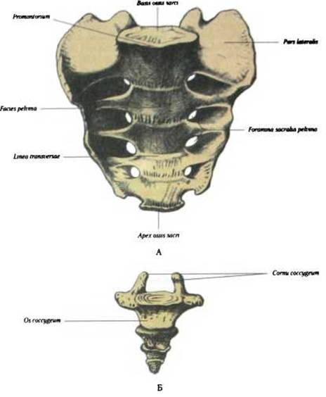
Fig. 30. Sacrum, os sacrum (A) and coccyx, os coccygis (B) — pelvic surface, facies pelvica
The apex of the sacrum is narrow and blunt, featuring a small facet for articulation with the coccyx to form the sacrococcygeal symphysis, art. sacrococcygea. On the dorsal surface of the sacrum, the intermediate crests terminate inferiorly in two small downward-pointing projections called the sacral cornua, cornua sacralia. The posterior surface of the apex and the sacral cornua bound the inferior opening of the sacral canal, known as the sacral hiatus, hiatus sacralis. The upper lateral region of the sacrum—the lateral part, pars lateralis—is formed by the fusion of the transverse processes of the sacral vertebrae. On the lateral surface of this region lies an ear-shaped articular surface, the auricular surface, facies auricularis, posterior and medial to which is a well-developed sacral tuberosity, tuberositas sacralis.

Fig. 31. Sacrum, os sacrum (A) and coccyx, os coccygis (B) — dorsal surface
Ossification. The primary ossification centers in the sacral vertebrae appear during the eighth month of intrauterine development. Fusion of the sacral vertebrae occurs gradually, beginning around 14–15 years of age and finishing between 17 and 25 years. Ossification starts in the lower vertebrae and progressively extends upward.
Last update: 08/08/2026
Editorial and Educational Adaptation: This material has been compiled based on the primary/original source text. The project team performed an editorial review, corrected technical inaccuracies, structured sections, and adapted the content for an educational format.
What was processed:
- elimination of formatting defects (OCR errors, structural breaks, corrupted characters);
- editorial organization of content;
- standardization of terminology in accordance with academic sources;
- verification of factual statements against the original source text.
All mentions of the author, publication year, and origin of the primary text have been preserved in accordance with the source.