Protein Structure and Function. Application of Bioinformatics Methods - John Rigden 2014
Prediction of protein function from its surface properties
Ligand-protein interaction
Annotation of ligand-binding sites
In drug discovery, the first critical step is predicting protein active sites and their ability to bind small molecules of hypothetical drug candidates. The second major step involves screening libraries containing tens of thousands of compounds to select those most likely to be selective and potent, ultimately leading to an effective therapeutic agent. This Selection process presents another challenge where an understanding of protein Structure/108.html">Surface Properties can also be helpful. Typically, these tasks are initially addressed using virtual Ligand screening, which incorporates molecular docking or pharmacophore searching Methods (Oledzki et al. 2006).
However, energetically driven annotations of ligand-binding sites can help visualize the most energetically significant surface points and thereby guide the application of computational methods toward them. Pioneering work in such energetic approaches was initiated by Peter Goodford with his widely used GRID program (Goodford 1985). In this method, as well as in subsequent ones, the interaction between a protein and a probe atom at a grid node on the protein surface is represented in terms of Van der Waals, hydrogen bonding, and Electrostatic Interactions. Particularly favorable interaction sites can be visualized (Fig. 7.5) and subsequently utilized in the design of novel ligands. Another similar approach involves applying knowledge-based potentials derived from existing data. For instance, the SuperStar program (Nissink et al. 2000) is based on a statistical analysis of numerous ligand-Protein Interactions extracted from structural Databases. Thus, both approaches can prove valuable in annotating potential active sites by identifying regions where favorable interactions between chemical groups are most likely to occur.
Class="center">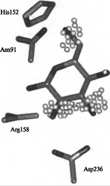
Fig. 7.5. Spatial grid generated by the Q-fit program (Jackson, 2002) for the Active Site of a periplasmic binding protein (PDB code 2GBP). The most favorable interaction points on the grid for oxygen hydroxyls are shown (highlighted in white). The bound ligand, glucose, was excluded from the calculations and is shown for comparison. As can be seen, four out of the five hydroxyl groups of glucose fall within the regions most favorable for probe atom placement.
Last update: 06/08/2026
Editorial and Educational Adaptation: This material has been compiled based on the primary/original source text. The project team performed an editorial review, corrected technical inaccuracies, structured sections, and adapted the content for an educational format.
What was processed:
- elimination of formatting defects (OCR errors, structural breaks, corrupted characters);
- editorial organization of content;
- standardization of terminology in accordance with academic sources;
- verification of factual statements against the original source text.
All mentions of the author, publication year, and origin of the primary text have been preserved in accordance with the source.