BIOCHEMISTRY - L. Stryer - 1984
VOLUME 1
PART I. CONFORMATION AND DYNAMICS
CHAPTER 8. ZYMOGEN ACTIVATION: DIGESTIVE ENZYMES AND CLOTTING FACTORS
8.27. The Extrinsic Pathway of Blood Coagulation
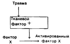
The extrinsic pathway of Blood Coagulation, like the intrinsic pathway, ultimately leads to the activation of Factor X. The extrinsic pathway (Fig. 8.38) is relatively simple. When a blood vessel is damaged, a lipoprotein called tissue factor is released. The complex of tissue factor and Factor VII catalyzes the activation of Factor X. Tissue factor Functions as a modifier protein, as Factor VII lacks activity in its absence. Thus, at least three modifier Proteins are involved in the blood clotting process: Factor V, Factor VIII, and tissue factor.
Class="center">Fig. 8.38. The extrinsic pathway of blood coagulation
8.28. Control of Blood Coagulation: A Problem Demanding Attention
It is quite obvious that blood coagulation must be very precisely regulated. The line between Hemorrhage and a tendency toward thrombosis is extremely fine. Clot formation must occur very rapidly, yet be strictly confined to the site of vessel injury. Why, then, does this process involve so many steps? Why do both intrinsic and extrinsic pathways exist, and how are they interconnected? What mechanisms limit the clotting process to the site of injury? These questions are fascinating not only in their own right but also in connection with the practical challenges of preventing and treating cardiovascular diseases.
The lability of activated clotting factors plays a crucial role in regulating clot formation. Their lifespan is very short because they are rapidly diluted in the bloodstream, cleared by the Liver, degraded by proteinases, and inactivated by specific inhibitors. The most important among these specific inhibitors is antithrombin III, a plasma protein that inactivates Thrombin by forming a stable complex with it. This complex appears to be similar to the stable acyl-enzyme complex formed by Trypsin and its pancreatic inhibitor (Section 8.12). Antithrombin III also inhibits clotting factors IXa, Xa, and XIa. The inhibitory action of antithrombin III is enhanced in the presence of heparin, a negatively charged polysaccharide (Section 9.16) synthesized by mast Cells located near the walls of Blood Vessels. Heparin acts as an anticoagulant by increasing The rate of irreversible complex formation between thrombin and antithrombin III.
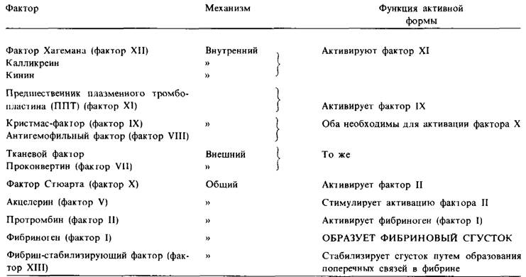
Table 8.2. Blood clotting factors
Last update: 06/08/2026
Editorial and Educational Adaptation: This material has been compiled based on the primary/original source text. The project team performed an editorial review, corrected technical inaccuracies, structured sections, and adapted the content for an educational format.
What was processed:
- elimination of formatting defects (OCR errors, structural breaks, corrupted characters);
- editorial organization of content;
- standardization of terminology in accordance with academic sources;
- verification of factual statements against the original source text.
All mentions of the author, publication year, and origin of the primary text have been preserved in accordance with the source.