Clinical and Morphological Diagnosis and Treatment of Sexually Transmitted Infections - Yakymova T. P. 2007
Appendix
Cervical smear in leukoplakia with atypia. In the center, a keratinized superficial epitheliocyte; to its right, a "naked" nucleus of an atypical cell and two macrophages; above, a malignant koilocyt. Pappenheim stain (x630)
Class="center">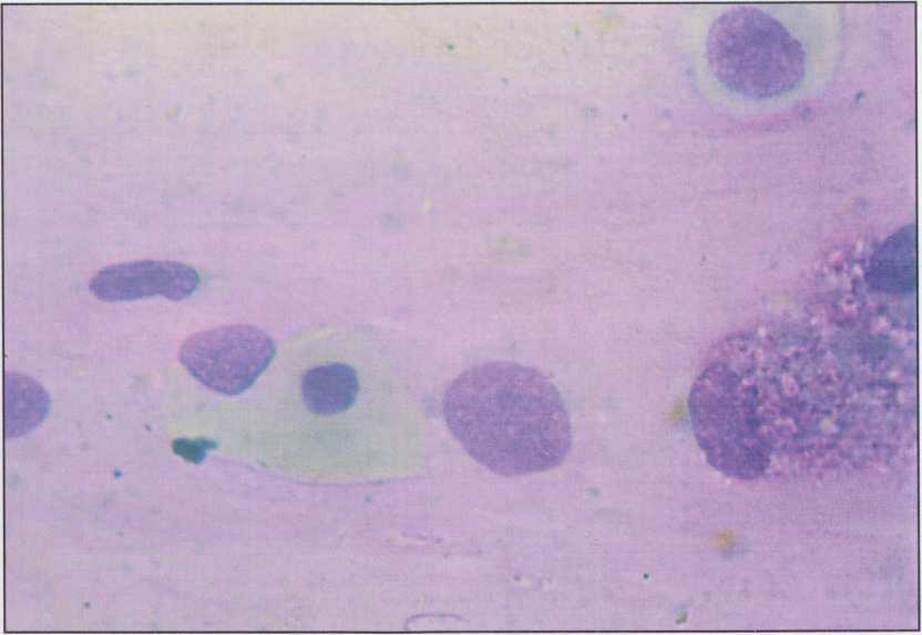
Fig. 58. Cervical smear in Leukoplakia with atypia. In the center, a keratinized superficial epitheliocyte is visible; to the right of it are a "naked" Nucleus of an atypical Cell and two macrophages, and at the top, a malignant koilocyte. Pappenheim stain (x630).
Last update: 13/08/2026
Editorial and Educational Adaptation: This material has been compiled based on the primary/original source text. The project team performed an editorial review, corrected technical inaccuracies, structured sections, and adapted the content for an educational format.
What was processed:
- elimination of formatting defects (OCR errors, structural breaks, corrupted characters);
- editorial organization of content;
- standardization of terminology in accordance with academic sources;
- verification of factual statements against the original source text.
All mentions of the author, publication year, and origin of the primary text have been preserved in accordance with the source.