Pathological Anatomy. Atlas of Macroscopic Specimens - Part 1. General Pathological Processes - A. M. Romaniuk 2016
Morphology of inflammation and immunopathological processes
Fibrinous-hemorrhagic cystitis
Class="center">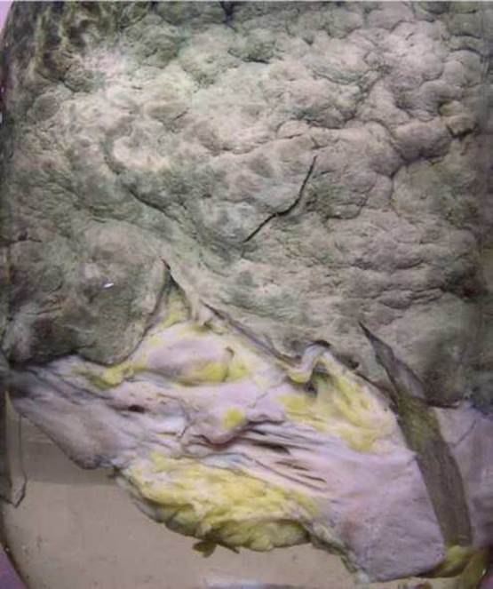
Fibrinous-hemorrhagic cystitis
The specimen shows the wall of the Urinary Bladder, with the mucous membrane exhibiting hypertrophy, fibrin deposition, and dark brown hemorrhages. This is an example of mixed exudative inflammation. Causes: infections; urinary stasis. Consequences: sclerotic changes.
Last update: 10/08/2026
Editorial and Educational Adaptation: This material has been compiled based on the primary/original source text. The project team performed an editorial review, corrected technical inaccuracies, structured sections, and adapted the content for an educational format.
What was processed:
- elimination of formatting defects (OCR errors, structural breaks, corrupted characters);
- editorial organization of content;
- standardization of terminology in accordance with academic sources;
- verification of factual statements against the original source text.
All mentions of the author, publication year, and origin of the primary text have been preserved in accordance with the source.