Fundamentals of Biochemistry (General Biological Introduction. Static Biochemistry) - I.V. Berezin - 1990
Fundamentals of Biochemistry (General Biological Introduction. Static Biochemistry) - I.V. Berezin - 1990
Preface
The last three to four decades have been characterized by the rapid development of biochemical research. Driven by the advancement of physical, chemical, and physicochemical methods for the isolation, purification, analysis, and study of naturally occurring substances, new scientific fields have emerged from classical biochemistry over this period: molecular biology, bioorganic and bioinorganic chemistry, biophysical chemistry, physical biochemistry, physicochemical biology, chemical enzymology, and other more specialized disciplines. Building upon their fundamental achievements, biotechnology has gained widespread recognition as an applied field.
Because modern biochemical research extensively employs physical and chemical methods and approaches, computer technology, and mathematical modeling, there is a growing demand for biochemical specialists with a profound background in chemistry, physics, and mathematics. This trend has actually been evident for quite some time. The need for a definite reorientation in the training of biochemists was highlighted as early as 20 years ago by Academician A.A. Baev and Prof. A.M. Varshavsky. In the preface to the renowned translated textbook "Fundamentals of Biochemistry" by H. Mahler and E. Cordes, they wrote: "Unfortunately, by tradition, biological chemistry is still classified among the biological disciplines, and consequently, specialists in biochemistry are trained in medical institutes and biological faculties. Meanwhile, in its modern form, biochemistry has long been essentially a branch of chemistry. Undoubtedly, a less severe disadvantage for a biochemist would be inadequate training in botany and zoology rather than a weak knowledge of mathematics, physics, and chemistry."
The necessity of teaching biochemistry as both a general education and a specialized subject in chemistry departments seems no longer open to dispute (it is worth noting, incidentally, that training biophysicists in both biological and physical faculties of universities has never been considered redundant duplication). For over a decade, the Department of Chemistry at Moscow State University (Chair of Chemical Enzymology) has been training specialists in enzymatic catalysis—one of the core areas of biochemistry. Although the level of their specialized knowledge is, judging by feedback, rated very highly, there has been a constant need to broaden their general educational training in biochemistry as a whole and to make it more systematic. To this end, a 54-hour lecture course was introduced a few years ago. It is delivered to fourth-year students after they complete their general training in chemistry, physics, and mathematics, but before they begin taking specialized elective courses. The objective of this introductory course is to help students acquire a sort of "foundational" yet broad-based education in all aspects of modern biochemistry, enabling them to easily adapt later to any narrower scientific field within the biochemical or even biomedical domain. A distinctive feature of this lecture course is that it is designed for students of chemical and other non-biological or non-medical faculties. Furthermore, grasping its material does not require advanced prior preparation in chemistry, physics, mathematics, or biology. An active proponent of this approach is the biochemist, scientist, and educator A. Lehninger. He believes that the time has come to include an introductory course in biochemistry in the curricula not only of biological and medical faculties, but also of chemical, physical, and other faculties in higher education institutions. A. Lehninger justifies his view by pointing to the position that contemporary biochemistry occupies in modern natural science, the role it plays in shaping the worldview of any educated person regardless of their field of activity, and the understanding it fosters regarding the importance of preserving living nature and human health.
The book offered to the readers contains material from only the first half of the aforementioned introductory biochemistry lecture course. It encompasses the general biological introduction to biochemistry and static biochemistry.
The general biological introduction familiarizes the reader with the structure and diversity of living nature at the cellular, tissue, organ, and organismal levels. It compiles and systematizes material that is typically little known to students in non-biological specialties, yet is indispensable for future biochemists.
Static biochemistry introduces the structure and properties of the principal substances of living nature at the molecular level. Wherever relevant, their functional purpose is noted. This section gathers and systematizes material that in general courses, even within chemistry departments, is presented in a highly fragmented manner. A sound understanding of the structure and properties of the primary chemical components of living nature will undoubtedly facilitate the subsequent mastery of biochemistry sections dedicated to biological processes.
Thus, the general biological introduction and static biochemistry can be regarded as the foundational "alphabet"—the bedrock for those who intend to master the "language" of biochemistry. In the future, it is planned to publish the material for the second half of the lecture course, which will include chapters on dynamic biochemistry and molecular biology, with the potential inclusion of functional biochemistry.
In form, this tutorial is a set of lecture notes. This means that the core information is presented in figures, while the text often serves as concise commentary on them. The authors hope that this textbook will prove useful to students in chemical and other faculties, as well as to graduates of these faculties whose research interests lie at the intersection of chemistry, physics, and biology.
In conclusion, the authors would like to express their gratitude to Professors N.N. Ugarova, A.K. Yatsimirsky, S.D. Varfolomeev, Doctor of Biological Sciences A.M. Egorov, and also to N.M. Glazkova, Head of Editorial at Moscow State University Press, for their assistance in working on the manuscript, which made its completion possible despite the sudden and grievous loss of Ilya Vasilyevich Berezin. We would also like to acknowledge K.I. Sautieva and V.V. Zaitsev for their meticulous technical preparation of the manuscript for publication.
Yu.V. SAVIN
Introduction
DEFINITION OF MODERN BIOCHEMISTRY
§ 1.
The Subject Matter of Biochemistry
Every science is defined by its object, methods, and objectives of study. From this perspective, what is biochemistry? Its very name merely indicates that it is somehow related to both biology and chemistry. But in what way?
Chemistry is the science that studies the composition, structure, properties, and transformations of natural substances for the purpose of obtaining them and synthesizing new substances to benefit humanity.
Biology is the science that studies the diversity of living organisms, their structure and functioning, and their interactions with the environment in order to better understand humanity and determine its place in the living world.
Since the definition of biology employs the term "living nature," it is necessary to clarify how it differs from the rest of nature, i.e., inanimate nature.
Distinguishing features of living nature:
structural complexity coupled with a remarkably small diversity of simple constituent components;
a high degree of internal organization with a strictly defined function for each constituent part;
the ability to maintain a living state by extracting energy and matter from the environment;
the ability to reproduce indefinitely.
Based on the definitions of chemistry and biology, it becomes clear how biochemistry relates to them. Biochemistry is chemistry because it studies the composition, structure, properties, and transformations of substances, and it becomes biological, first, because it investigates not just any substances, but only those found and undergoing various transformations in living nature, and second, because this study aims to understand the functioning of living objects.
Thus, biochemistry is a science that studies the composition, structure, and properties of substances in living nature, as well as their transformations in the course of the vital activity of living organisms, in order to uncover the molecular foundations of life. Solving this ultimate task drives progress in fields related to human healthcare and living conditions, and ultimately provides an answer to the fundamental question of natural science regarding the origin of life.
§ 2.
Modern Biochemistry: A Qualitatively
and Quantitatively New Stage
in the Development of Biochemistry
The development of any science proceeds in leaps and bounds. The period of quantitative accumulation of facts culminates at a certain stage in the discovery of a series of general regularities and the creation of generalizing theories, which form a qualitatively new foundation for a subsequent round of quantitative fact accumulation.
For instance, three periods are recognized in the development of chemistry. Alchemy represents the pre-scientific period of gathering experimental data from ancient times up to the 18th century. Modern chemistry emerged in the mid-18th century with the discovery of the first quantitative laws and the establishment of atomic theory in the works of Lomonosov, Lavoisier, and Dalton. Contemporary chemistry began with D. I. Mendeleev's discovery of the periodic law, which systematized the knowledge accumulated in the preceding period and allowed, from a qualitatively new perspective, not only to explain experimental data but also to predict new ones.
Three periods can also be distinguished in the development of biochemistry.
Pre-scientific biochemistry is the period of accumulating practical knowledge (bread baking, cheese making, wine and beer production, tanning, etc.) from ancient times to the mid-19th century.
Classical biochemistry emerged as an independent science from physiology in the second half of the 19th century. Its inception was driven by the understandable desire to explain physiological processes through chemical reactions. However, how could biochemists approach this chemical task when for many decades they had to work not with pure substances, but with tissue suspensions, tissue fluids, and so on? For this reason, classical biochemistry is not unjustly classified among chemical sciences.
Modern biochemistry arose on the basis of classical biochemistry in the second half of the 19th century with the transition of biochemical research to a qualitatively new level—the molecular level. This became possible thanks to the advancement of chemical, physical, and physicochemical methods for the isolation, purification, analysis, and study of biological substances by that time. A qualitatively new level of research entailed a quantitative expansion of biochemical fields. Thus, on the basis of classical biochemistry, molecular biology, bioorganic and bioinorganic chemistry, biophysical chemistry and physical biochemistry, physicochemical biology, chemical enzymology, and others emerged. They differ from one another only in their research methods and approaches, as well as the choice of specific biological objects, yet they all pursue the same core goal of biochemistry: understanding the chemical foundations of life processes. This qualitatively new molecular—i.e., chemical—level of research allows modern biochemistry to be classified strictly as a chemical science.
Part I
GENERAL BIOLOGICAL INTRODUCTION
§ 1.
Classification of Living Objects
by the Nature of Their Interaction
with the Environment
As seen from the definition of life given above, the functioning of living objects is sustained by the uptake from the external environment of, first, matter, from the atoms of which (C, H, N, S, P, etc.) they synthesize their own structures, and second, energy, which drives this biosynthesis. In this regard, all living organisms are conventionally divided (Fig. 1.1)
by the type of matter utilized into:
lithotrophs (autotrophs), which consume simple inorganic compounds such as СО2, Н2О, N2, NH3, H2S, H2PO4-, and others, and organotrophs (heterotrophs), which consume various organic compounds;
by the type of energy utilized into:
phototrophs, which use light energy, and chemotrophs, which use the chemical bond energy of matter derived from the external environment;
in relation to atmospheric oxygen into:
aerobes, which use oxygen as an oxidizing agent and perish in its absence; facultative anaerobes, which can use oxygen as an oxidizing agent but do not die in its absence; and obligate anaerobes, for whom oxygen acts as a metabolic poison.
In photolithotrophs, such as green plants, algae, and cyanobacteria, oxygen is released during photosynthesis:
nН2О +nСО2
(СН2О)n+nО2. |
(I.1) |
In other photolithotrophs, specifically purple sulfur bacteria, photosynthesis proceeds without the release of oxygen:
2nH2S +nCO2
(CH2O)n+ 2nS +nH2O. |
(I.2) |

Fig. 1.1. Classification of living objects by the nature of their interaction with the environment
Chemolithotrophs are certain types of bacteria, such as nitrifying bacteria (aerobes), which assimilate СО2 utilizing energy from oxygen-driven oxidation reactions:
2NH4++ 5O2→ 2NO3-+ 4H2O, |
(I.3) |
or denitrifying bacteria (anaerobes) that assimilate СО2 using energy derived from nitrate reduction:
10S + 12NO3-+ 4H2O → 10SO42-+ 6N2+ 8H+. |
(I.4) |
Organotrophs (heterotrophs) are subdivided into photo- and chemoorganotrophs.
Photoorganotrophs, such as non-sulfur purple bacteria, require not only CO2 but also simple organic compounds, such as ethyl or isopropyl alcohols, as a carbon source:
nCH3CH2OH + nСО2
(СН2О)n + nСН3СООН. |
(I.5) |
Chemoorganotrophs are the principal representatives of organotrophs (heterotrophs). They obtain their nutrition at the expense of lithotrophs (autotrophs) or other organotrophs (heterotrophs).
Depending on their mode of nutrition, chemoorganotrophs are, in turn, subdivided into saprotrophs—organisms that feed on the remains of other organisms or their metabolic products (most fungi and many types of bacteria); parasites—organisms that live on the surface or inside other organisms, feeding at their expense (some fungi, many types of bacteria, certain plants and animals); and holozoic organisms—most animals that actively seek out and consume other organisms.
Section A
THE CELL
The cell is the smallest structural and functional unit of living matter. Like any living matter, a cell consists of molecules of various substances that interact according to the laws of chemistry and physics; however, it is only within the cell that this interaction results in the manifestation of the characteristics of living matter. The minimum size of a cell is limited by the dimensions of its constituent molecules, whereas its maximum size is determined by the laws governing the diffusion of substances into and within the cell. Since a cell's surface area increases in proportion to the square of its radius as it grows, while its volume increases in proportion to the cube of its radius, beyond a certain size the cell will experience a shortage of nutrients and become nonviable.
Based on structural and functional features, all cells are divided into prokaryotic ("pre-nuclear") and eukaryotic (with a well-formed nucleus). In nature, prokaryotic cells are represented by organisms called prokaryotes, and eukaryotic cells by eukaryotes.
Chapter 1
PROKARYOTIC CELLS
§ 1.
Distribution,
shapes, and sizes
In the living world, prokaryotic cells are represented exclusively by bacteria—the majority of microorganisms, which account for three-quarters of living matter on Earth. Bacteria come in various shapes: rod-shaped, or bacilli (bacillus); spherical, or cocci (coccus), which are referred to as gonococci and diplococci respectively if they occur singly or in pairs, as streptococci if arranged in chains, and as staphylococci if clustered in grapnel-like bunches; comma-shaped bacteria, or vibrios (vibrio), such as the cholera bacterium; slightly twisted spirals, or spirilla (spirillum); and tightly coiled spirals, or spirochetes (spirochaetales).
The typical dimensions of bacteria range from single digits to tens of micrometers, though some bacteria can reach lengths of up to hundreds of micrometers, such as spirochetes (up to 500 µm), a group that includes the causative agent of syphilis.
§ 2.
Structure
The structure of a prokaryotic cell is illustrated in Fig. 1.2.
The cell is externally enclosed by a cell envelope that separates its internal contents from the external environment. The cell envelope has the ability to allow essential nutrients to pass inward, while

Fig. I.2. Structure of a prokaryotic cell
allowing metabolic waste products to pass outward. All bacteria are classified as either gram-positive or gram-negative based on their ability to retain or not retain the Gram stain (using successive treatments with crystal violet, iodine, and safranin). This division is associated with two distinct types of cell wall architecture.
In gram-positive bacteria, the envelope consists of a plasma membrane—composed of lipids and proteins—and a rigid, thick cell wall overlying it, the structural basis of which is polysaccharides cross-linked by short peptides. In gram-negative bacteria, the plasma membrane is covered by a thin polysaccharide cell wall, which in turn is protected by an outer membrane composed of lipids, proteins, and lipopolysaccharides. Across all cells, the plasma membrane has a nearly uniform thickness and identical structure (see Section "Lipids"). Its distinctive feature is that it contains a large number of associated enzymes that provide the cell with chemical energy in the form of ATP derived from external nutrients or light; drive the directed transport of substances across the membrane utilizing ATP energy; and facilitate the synthesis of the cell wall.
The cell wall maintains the shape of bacteria and protects them from osmotic lysis.
Protoplasts and spheroplasts are gram-positive and gram-negative bacteria, respectively, that have been artificially stripped of their cell walls and maintained in a hypertonic medium to preserve their integrity.
Mesosomes are multilaminar structures formed by invaginations of the plasma membrane. They are not present in all bacteria. Membrane-bound enzymes of mesosomes participate in numerous metabolic reactions, particularly in DNA replication.
Cytoplasm is the internal medium of the cell where the majority of metabolic processes take place.
Cytosol is the liquid portion of the cytoplasm.
Ribosomes are granules with a maximum diameter of about 20 nm, composed of RNA and proteins. The synthesis of cellular proteins takes place on ribosomes.
Polysomes, or polyribosomes, are strands of messenger RNA with ribosomes strung along them like beads.
The nucleoid (or nuclear body) is a densely packed circular double-stranded DNA molecule. This single "chromosome" carries the cell's core genetic information and is not separated from the cytoplasm by a membrane.
Plasmids are very small, extrachromosomal circular DNA molecules located in the cytoplasm. They encode essential information that determines cell survival under specific conditions, notably the structure of enzymes that neutralize certain antibiotics.
Episomes are plasmids capable of integrating into the chromosomal DNA under certain conditions. Episomes that can leave chromosomes while carrying along fragments of chromosomal DNA are referred to as sex factors (F factors).
Pili (fimbriae) are thin, straight, hollow filaments covering the cell wall. DNA is transferred from one bacterium to another through sex pili (F-pili).
Flagella are long filaments, thicker than pili and often clustered in bundles. Their rotation causes the bacterium to reverse direction and propel forward. Bacterial flagellar rotation is driven by a unique "proton motor" located in the plasma membrane.
Various granules containing stored reserves may also be found in the bacterial cytoplasm. These can include granules of poly-β-hydroxybutyric acid, glycogen, polyphosphate, protein, or lipid droplets.
Vacuoles are droplets of an isolated aqueous phase containing dissolved substances.
Thus, a primitive division of labor exists in the prokaryotic cell: the cell wall provides protection, the plasma membrane facilitates nutrient uptake and the extraction of chemical energy therefrom, polyribosomes synthesize proteins, and the nucleoid and plasmids store and transmit genetic information, etc.
§ 3.
Reproduction
Bacterial reproduction typically occurs through simple binary fission. Prior to division, the DNA content in the nuclear body doubles, and the cell then divides such that the daughter cells receive DNA identical to the parental DNA prior to replication. Under certain conditions, some bacterial species are also capable of reproducing via conjugation, a process involving either the fusion of two cells or the transfer of a single DNA strand through a pilus from one bacterium to another. Subsequent exchange of genetic material occurs within the cell, and following division, the daughter cells receive DNA carrying information derived from both parent cells.
§ 4.
Diversity
The diversity of prokaryotic cells is reflected in the vast array of bacteria, which comprise about 3,000 known species. Bacteria are conventionally divided into 20 groups. Their classification is based on a combination of multiple features: shape, Gram-staining characteristics, mode of nutrition, spore-forming ability, production of specific metabolites, oxygen requirements, etc.
Four groups of bacteria deserve special mention.
Cyanobacteria, also referred to as blue-green algae, are the only photosynthetic bacteria capable of releasing molecular oxygen.
Rickettsiae are primitive bacteria with very modest nutritional requirements, capable of living and reproducing exclusively inside host cells.
Mycoplasmas are distinguished by their extremely small size (about 300 nm in diameter) and the absence of a rigid cell wall. By altering their shape, they can squeeze through pores smaller than their own diameter. They exist solely as parasites within other organisms.
Spore-forming bacteria produce spores under unfavorable environmental conditions—microcells containing essential reserves of nutrients and genetic material, featuring a tough protective coat and virtually complete dehydration. Once the parent cell disintegrates, a spore can persist in adverse conditions indefinitely. However, upon encountering a favorable environment, it germinates back into a normal bacterium.
Recently, it has become standard practice to segregate methanogenic bacteria (which produce methane during metabolism), halobacteria (which inhabit environments with exceptionally high salt concentrations), and thermoacidophiles (which thrive in acidic environments at extremely high temperatures) into a distinct group known as archaebacteria. This classification is driven by their unique molecular architecture and biochemical functioning. All other bacteria are referred to as true bacteria, or eubacteria.
Chapter 2
EUKARYOTIC CELLS
§ 1.
Distribution,
Shapes, and Sizes
Eukaryotic cells comprise the cells of all organisms except bacteria: single-celled protozoa, fungi, algae, plants, and animals. Compared to prokaryotes, eukaryotes are far more abundant, with their species numbering in the millions.
Eukaryotic cells exhibit remarkable diversity in shape and size. Some are enormous, such as the chicken egg cell (yolk), or exceptionally long, such as certain nerve cells with axons in squids that reach several meters in length. Overall, eukaryotic cells are significantly larger and more complex than prokaryotic cells. They typically have diameters measured in tens of micrometers and volumes several thousand times greater than those of prokaryotes.
§ 2.
Structure
Eukaryotic cells are characterized by a well-defined nucleus and a rich array of subcellular organelles; some cells are multinucleated. Fig. 1.3 illustrates a "generalized" eukaryotic cell. Let us examine its subcellular structures, moving from the center toward the periphery.
The nucleus is separated from the rest of the cell contents by the nuclear envelope, which consists of an outer and an inner membrane with a 20–40 nm perinuclear space between them. The structure of the nuclear membranes is similar to that of the plasma membrane. The nuclear envelope features numerous pores (up to 100 nm in diameter). Chromatin, the nuclear content, is named for its affinity for specific stains. It is a mixture of DNA (35%), RNA (5%), and proteins (60%). When the cell is not dividing, chromatin is distributed uniformly throughout the nuclear sap (nucleoplasm) and appears as a network of chromatin threads visible only under an electron microscope. At the onset of cell division, these chromatin threads coil into supercoils to form dense structures known as chromosomes, which can be seen under a standard light microscope. Fig. 1.4 shows the human chromosome set. The nucleus is the site of DNA and RNA synthesis.
The nucleolus is a denser and more intensely staining structure within the nucleus compared to chromatin. It disappears during cell division and contains the bulk of nuclear RNA. It is the site of ribosome assembly. A nucleus may contain several nucleoli.
The eukaryotic cytoplasm has a finer structure than the prokaryotic one. In addition to the liquid phase—the cytosol—it contains a cytoskeleton, which is a dense network of microfilaments, myofiblaments, and microtubules. It helps maintain cell shape, particularly in animal cells, which have rather pliable envelopes, and ensures the relative positioning of numerous subcellular structures (organelles).
The endoplasmic reticulum (ER) is a complex, branched, three-dimensional network of channels and cavities permeating the entire cytoplasm. The walls of these channels and cavities (cisternae) have a membranous structure similar to the plasma membrane. Rough ER differs from smooth ER by the presence of ribosomes on the outer surface of the membranes that form its channels and cisternae. The primary function of the rough ER is protein synthesis, whereas carbohydrates and lipids are synthesized on the outer surface of smooth ER membranes. The synthesized proteins, carbohydrates, and lipids cross the ER membranes to their inner side and are transported through the ER channels to their destinations. The cisternae act as storage reservoirs for these synthesized substances.
Eukaryotic ribosomes are slightly larger than prokaryotic ones. They consist of RNA-protein complexes and, like those in prokaryotic cells, lack a membrane.

Fig. I.3. Structure of a eukaryotic cell
The Golgi apparatus is a stack of flat cavities and tubules formed from single membranes. Along the edges, individual vesicles can be seen budding off from the main body. The Golgi apparatus is typically located
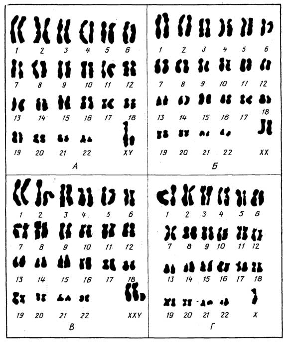
Fig. I.4. Human chromosome set: male (A), female (B), genetic abnormalities (C, D)
not far from the nucleus, though in some cells, such as nerve cells, it completely surrounds it. In plant cells, it is referred to as a dictyosome. Compounds synthesized in the ER first enter the Golgi apparatus, where they are stored and modified, and then transported in vesicles to their cellular destinations or secreted outside the cell (exocytosis). The membranes of the Golgi apparatus also synthesize their own lipids and carbohydrates. The Golgi apparatus is absent in erythrocytes and spermatozoa.
Lysosomes are small vesicles bounded by a single membrane that contain enzymes responsible for breaking down cellular proteins, lipids, and carbohydrates that are either foreign or no longer needed by the cell.
Microbodies are organelles resembling lysosomes, though they frequently differ by the presence of an internal dense core consisting of protein crystals. Peroxisomes are microbodies found in animal and plant cells that contain a group of enzymes involved in the production or consumption of hydrogen peroxide. Glyoxysomes are plant cell microbodies containing enzymes of the glyoxylate cycle, which enable the synthesis of carbohydrates from lipid fatty acids.
Vacuoles are single-membrane vesicles containing various metabolic products. In young plant cells, they are small, but they increase significantly in size as the cell ages. They are rarely found in animal cells.
Granules serve as nutrient reserves. They contain solid forms of proteins, polysaccharides (glycogen, starch), and lipids.
Fat droplets represent a mechanism for storing liquid lipids.
Mitochondria are relatively large double-membrane organelles with a complex internal structure formed by numerous folds (cristae) of the inner membrane. Mitochondria contain enzymes capable of converting the chemical bond energy of nutrients into the universal energy carrier ATP (see the section "Nucleotides") using an oxidizing agent (O2). Mitochondria also contain a small circular DNA molecule and ribosomes similar to those of bacteria. During cell division, mitochondria replicate as well.
Plastids are plant cell organelles significantly larger than mitochondria, yet possessing both outer and inner membranes like the latter. They also divide during cell division. Plastids are classified into three types: chloroplasts, chromoplasts, and leucoplasts, all of which are capable of interconversion. Chloroplasts are plastids containing chlorophyll as their primary pigment and are capable of photosynthesis. Inside chloroplasts, the inner membrane forms disc-like thylakoid vesicles distributed throughout the organelle and grouped into grana. Chloroplasts are located in the green tissues of plants. Like mitochondria, they contain DNA. Chromoplasts, unlike chloroplasts, contain other pigments from the carotenoid group in addition to chlorophyll (see the section "Lipids"). The presence of chromoplasts accounts for the yellow, orange, and red colors of flowers, fruits, and autumn leaves. Leucoplasts are colorless plastids in which plant cells store starch and oils.
The centrosome plays a crucial role in organizing chromosome division. In animal cells, it is represented by two small bodies known as centrioles.
The cell envelope separates the contents of a eukaryotic cell from its external environment. In plant cells, it consists of a plasma membrane protected externally by a rigid cell wall. In animal cells, the cell envelope consists solely of a pliable plasma membrane.
The cell wall of plant cells determines cell shape and provides structural support and protection. Its structural foundation consists of cellulose microfibrils cemented together by a mixture of polysaccharides, proteins, and a highly branched polymer, lignin, which contains aromatic residues in its chains (see the section "Carbohydrates");
The plasma membrane regulates the exchange of substances between the cell interior and the external environment. In all cells, it is composed of lipids and proteins. The plasma membrane occasionally forms folds that extend deep into the cell, reaching all the way to the nuclear envelope and connecting with it. The ability of certain unicellular microorganisms, such as amoebas, as well as specific animal cells, such as leukocytes, to ingest relatively large nutrient particles—whether solid (phagocytosis) or liquid (pinocytosis)—is due to the properties of the plasma membrane. This process occurs as follows: the membrane forms an invagination around the "adhering" particle, after which its edges fuse together.
Microsomes are small vesicles (about 100 nm in diameter) formed from the membranes of the ER and/or the plasma membrane. Ribosomes may be present on their outer surface. Microsomes are formed following cell disruption and are isolated by centrifugation; they are not found in living cells.
Microvilli are small, slender projections of the plasma membrane in animal cells that significantly increase their surface area.
Cilia are structures longer and thicker than microvilli. Their internal architecture enables rotational movement powered by ATP hydrolysis. Their function is to propel substances along the cell surface or to drive the locomotion of the cell itself.
Flagella have a similar structure, but they are several times longer. By rotating, flagella enable single cells to move rapidly.
§ 3.
Formation of Multicellular Structures
Eukaryotic cells differ from prokaryotic cells in their ability to interact with one another and form complex multicellular structures—tissues, organs, and organisms.
Eukaryotic cells are held together in various ways.
In the absence of direct contact, this is achieved 1) through the adhesive action of the extracellular matrix, which contains proteins. Therefore, when proteins are degraded, for example enzymatically, the cells separate.
Semi-direct contact occurs through 2) the formation of desmosomes (junctional complexes)—regularly arranged, numerous membrane partitions between loosely spaced plasma membranes of neighboring cells; and 3) via short or long flexible cell extensions (filaments) through which cells can exchange cytoplasmic contents.
Direct contact is achieved through the formation of tight junctions, which may involve 4) the interlocking of cell walls via mutually penetrating invaginations and evaginations; 5) the simple fusion of plasma membranes at contact sites; 6) the formation of gap junctions, i.e., sufficiently large pores in contiguous cell walls; and 7) the formation of plasmodesmata—membrane tubules that pierce the rigid walls of plant cells and facilitate cytoplasmic exchange between them.
Special forms of eukaryotic cell aggregation in multicellular organisms include syncytia and symplasts, where cells partially or completely fuse. Syncytia (or coenocytes) represent an integration of cells into a single entity connected by cytoplasmic extensions (filaments) or gap junctions, allowing them to exchange cytoplasmic contents relatively freely while their nuclei retain functional independence. In symplasts, the boundaries between individual cells virtually disappear, forming a unified structure (or a giant cell) in which multiple nuclei reside within a common cytoplasm.
§ 4.
Reproduction
Based on their mode of reproduction, eukaryotic cells can be divided into two groups. The first group includes all cells except those responsible for reproducing the organism as a whole. In plants, these are called vegetative cells; in animals, somatic cells. They are formed through mitosis, a process in which a daughter cell receives the same amount of genetic material as the parent cell. Occasionally, cell reproduction occurs via simple division (amitosis). The second group consists of specialized cells responsible for reproducing the organism as a whole. These are germ cells. They are formed through meiosis—a process
Fig. I.5. Mitosis: interphase (A), prophase (B), metaphase (C), anaphase (D), telophase (E)
in which the daughter cell receives half as much genetic material as the parent cell.
Mitosis. This is a continuous process that can be conventionally divided into four stages (Fig. I.5): prophase, metaphase, anaphase, and telophase. In most cells, mitosis takes 0.5–3 hours, whereas the preparation for it (interphase) takes considerably longer (10–30 hours).
Interphase. Genetic material is doubled through DNA replication. Proteins involved in cell division are synthesized. ATP is synthesized, without which energy reserves cell division is impossible. The number of certain subcellular structures, such as mitochondria and chloroplasts, increases.
Prophase. The nucleus increases in size. Chromatin threads, each having undergone replication during interphase, form supercoils and transform into chromosomes visible under a standard microscope. Each chromosome consists of two interconnected chromatids (the result of replication). The point of attachment is called the centromere. Next, poles form within the cell, which essentially guide further division. In animal cells, the centriole divides, and daughter centrioles migrate to opposite poles of the cell. In plant cells, poles form without centrioles. Microtubules (15 nm)—fibers of the mitotic spindle—then extend from the poles toward the equator, with some attaching to the centromeres of the chromosomes. The nuclear envelope and nucleolus disappear.
Metaphase. Chromosomes align along the equator. This stage proceeds more rapidly than prophase.
Anaphase. Chromosomes separate at the centromeres into chromatids, which migrate to opposite poles.
Telophase. A nuclear envelope forms around the chromatids (now referred to as daughter chromosomes) that have gathered at the poles. The animal cell divides in half. In plant cells, a plasma membrane forms in the middle, dividing the parent cell into two daughter cells. In the newly formed cells, the chromatids (daughter chromosomes) revert to chromatin, and a nucleolus reappears.
Amitosis. This is a process of cell reproduction through simple division, where neither chromosome formation nor a mitotic spindle is observed, and the nuclear envelope and nucleolus do not disappear. Amitosis involves a simple division of the nucleus in half, followed by cytoplasmic division. Amitosis frequently does not result in complete cell division, leading instead to the formation of a binucleated cell. It has been observed in plant and animal cells, as well as in simpler organisms. Amitosis can be considered an atypical mode of reproduction, as daughter cells subsequently lose viability and divide no further.
Meiosis is the process of forming male and female germ cells (Fig. I.6). It comprises two divisions, each of

Fig. I.6. Meiosis. First division: prophase (A), metaphase (B), anaphase and telophase (C). Second division: prophase and metaphase (D), anaphase (E), telophase (F)
which can also be subdivided into prophase, metaphase, anaphase, and telophase.
Interphase. The same processes occur as in mitosis, i.e., DNA replication and the synthesis of essential proteins and ATP.
First Division
Prophase. Chromosomes form from chromatin just as in mitosis; however, unlike mitosis, chromosomes pair up because eukaryotic cells, other than germ cells, typically contain an even number of chromosomes. Chromosomes that make up such pairs are called homologous chromosomes. They carry similar genetic information, such as eye color, flower shape, etc. Furthermore, with the exception of the sex chromosomes which form only a single pair, they are identical in external characteristics (shape and structure); therefore, even in cells undergoing mitosis, all chromosomes can be grouped into homologous pairs. If a cell contains pairs of homologous chromosomes, it possesses a diploid set of chromosomes. If a cell contains a single homologous chromosome from each pair, it possesses a haploid set of chromosomes. Homologous chromosomes within pairs approach each other closely and intertwine by their chromatids, forming tetrads.
Between them, crossing over (or conjugation) takes place, during which the chromatid of one homologous chromosome exchanges genetic material (i. e., DNA) with a chromatid of the other homologous chromosome across certain regions. Since one homologous chromosome carries paternal information and the other maternal, following crossing over, each chromatid within the tetrad contains genetic information from both parents. Simultaneously, the cell poles and the spindle apparatus begin to form.
Metaphase. The conjugated homologous chromosomes align in pairs (as tetrads) along the equatorial plane.
Anaphase. The conjugated homologous chromosomes separate and migrate toward opposite poles. Each homologous chromosome still consists of two chromatids.
Telophase does not reach completion because the chromosomes do not decondense into chromatin, and no nucleolus forms. Instead, the newly emerging cells immediately proceed to the second division.
Second Division
Prophase. In each of the two newly formed cells, poles reappear (at a 90° angle relative to the previous ones), and a new spindle apparatus forms between them.
Metaphase. The chromosomes align along a new equator (perpendicular to the equator of the previous division).
Anaphase. As in mitosis, each chromosome divides into chromatids, which migrate to opposite poles.
Telophase. A nuclear envelope forms around each group of chromatids (now referred to as daughter chromosomes). The cell undergoes final division. In the resulting four new cells, the chromosomes decondense into chromatin, and nucleoli reappear.
Thus, in mitosis, following a single division, each of the two daughter cells retains the same number of chromosomes as the parent cell, whereas in meiosis, following two consecutive divisions, each of the four daughter cells receives half the chromosome number of the parent cell.
In both cases, the parent cell contains a diploid set of double chromosomes prior to division, while the daughter cells contain single chromosomes after division. However, following mitosis, these constitute a diploid set, whereas following meiosis, they form a haploid set.
Modes of reproduction
in eukaryotic organisms
Based on their modes of reproduction, eukaryotes can be divided into three groups.
The first group comprises organisms that reproduce asexually—that is, by division or budding—where one or more ordinary cells, previously detached from the parent and containing either a diploid or haploid set of chromosomes, give rise to a new organism through mitotic division. This type of reproduction is exclusive to certain simple unicellular organisms, such as amoebas.
The second group includes organisms that reproduce sexually using specialized germ cells containing a haploid set of chromosomes. The fusion of two such cells results in fertilization and the formation of a diploid zygote, which develops into a new organism. This mode of reproduction is characteristic of higher animals and plants.
There are also organisms capable of reproducing both asexually and sexually. This group includes many primitive organisms, lower animals, and numerous higher plants. The latter can reproduce vegetatively, for instance, via cuttings, grafting, or tubers.
§ 5.
Diversity
The diversity of eukaryotic cells is determined, firstly, by the vast variety of eukaryotic organisms, represented by millions of species, and, secondly, by cellular differentiation into specialized tissues in more complex organisms.
Since ancient times, the living world has been divided into plants and animals.
Differences between plants
and animals
Mode of nutrition: predominantly photolithotrophic in plants and chemoorganotrophic in animals.
Motility: plants are sessile or passively carried along with their habitat, whereas animals are capable of active locomotion (with rare exceptions among certain lower animals, such as tunicates, which lead a sessile lifestyle in their adult stage).
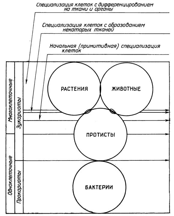
Fig. I.7. Principles of living organism classification
Presence of a cellulosic cell wall in plant cells and its absence in animal cells (with tunicates again serving as an exception).
Storage form of polysaccharides: starch in plants and glycogen in animals.
The capacity for continuous growth (increase in size and volume). Plants grow continuously throughout their lives. Animals, with few exceptions, grow to a certain size, after which only the replacement of old cells by new ones takes place.
Ever since the microbial world became accessible to study, many of its representatives could be classified as either plants or animals. Currently, eukaryotic organisms are generally divided into three kingdoms (Fig. I.7): Protista (protists), Metaphyta (plants), and Metazoa (animals). Each kingdom is divided into phyla and further into subphyla, classes, subclasses, orders, families, genera, and species. Protists can be unicellular or multicellular, whereas plants and animals are exclusively multicellular. The fundamental difference between protists, on the one hand, and plants and animals, on the other, lies in the absence of the key aromorphosis feature in the former—the formation of tissues and organs.
A tissue is an association of specialized cells that are similar in structure and function.
An organ is a part of a plant or animal organism that performs a specific function and may consist of various tissues. The emergence of organs is the result of further cell specialization.
"Division of labor among cells" associated with the formation of tissues and organs allows cells to function more efficiently, but at the same time increases the dependence of one cell type upon another.
Section B
EUKARYOTIC ORGANISMS.
STRUCTURAL FEATURES
AND VITAL ACTIVITY
Chapter 3
PROTISTS
This kingdom comprises primitive unicellular and multicellular organisms that often exhibit features of both plants and animals (Fig. I.8). In multicellular protists, all cells are identical, or if they differ, they do not group together to form tissues. Any initial specialization occurs strictly at the individual cell level.
§ 1.
Unicellular
protozoan organisms
Unicellular protists are listed here in order of decreasing animal traits and increasing plant traits.
Sarcodines are amoebas that lack a rigid cell wall, move by means of plasma membrane extensions (pseudopodia) and the flow of cytoplasm from one part of the cell to another, and feed via phagocytosis. Leukocytes in animals and humans behave similarly; foraminifera are also amoebas, but possess a perforated outer shell.
Sporozoans, represented by Plasmodium—a parasitic organism that causes malaria and is transported by the bloodstream.
Ciliates are of interest because they move by the beating of numerous cilia on the cell surface. They possess a primitive mouth, pharynx, and anal pore.
Flagellates form a large group of protozoa whose representatives possess features of both plants and animals. All of them actively move using one, two, or more flagella. Among flagellates, hemoflagellates and dinoflagellates are distinguished, as well as euglenids and chlamydomonads.
Hemoflagellates are microorganisms that clearly display animal characteristics. For instance, the single-flagellated trypanosomes prefer nervous tissue as their habitat. They are the causative agents of sleeping sickness. Trichonymphs, which inhabit the guts of termites, harbor bacteria capable of converting cellulose into glucose. These symbiotic bacteria provide nourishment not only for themselves but also for the trichonymphs, enabling termites to feed on wood.
Dinoflagellates are components of marine plankton. They possess a rigid shell and two flagella, yet, much like plants, they contain chlorophyll. Another pigment, fucoxanthin, imparts a red color to them. The presence of chlorophyll and fucoxanthin enables these organisms to lead a photolithotrophic lifestyle. However, pigment-free forms with chemoorganotrophic nutrition also occur among dinoflagellates.
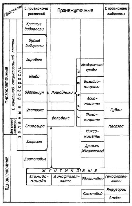
Fig. I.8. Classification of protists
Euglenids, possessing chloroplasts, are capable of photosynthesis; however, this does not fully meet the metabolic needs of the cells, and therefore euglenizing cultivation is only possible if amino acids are present in the nutrient medium. In the dark, they adopt an animal-like lifestyle by engulfing food particles, or detritus.
Using their flagellum, euglenids navigate toward well-lit areas, which they can detect thanks to a specialized organelle—the eyespot (stigma)—containing the light-sensitive pigment astaxanthin. Photosynthesis is not strictly obligatory for them: if artificially deprived of chloroplasts, they continue to survive as animals. Euglenids lack a cellulose wall. Their reserve substances are stored as the polysaccharide paramylon, which differs from both starch and glycogen.
Chlamydomonads are in many ways similar to euglenids. They also exhibit animal characteristics: a light-sensitive eyespot and two flagella that provide locomotion. Their plant-like traits include a single chloroplast that provides photosynthesis for essential substances, a robust cellulose cell wall, and energy storage in the form of starch.
Diatoms resemble plants more than animals. Often referred to as diatomaceous algae, together with dinoflagellates they form a major component of marine plankton, synthesizing approximately 75% of all organic matter on Earth. The diatom cell is encrusted with a rigid cell wall made of silicon dioxide (silica). Diatoms store reserve substances not as glycogen or starch, but as oil droplets. Fossilized deposits of their cell wall remnants are known as diatomaceous earth, while ancient diatom oil deposits may have transformed into petroleum reserves.
Green algae (Chlorophyta), which include the well-known Chlorella, already possess all the characteristic features of plants: chloroplasts, a rigid cellulose wall, and the accumulation of starch. Animal cell traits are entirely absent. Water "blooming" in ponds is primarily caused by accumulations of green algae.
Yeasts are fungi. They exhibit virtually all plant characteristics except for the presence of chlorophyll. They exist as unicellular saprophytes in environments with a relatively high sugar content.
§ 2.
Multicellular Protists
Among multicellular protists, there are organisms either lacking cell specialization or exhibiting only rudimentary cell-level specialization.
An example of animal-like organisms with virtually no cell specialization is found in the microscopic worms of the phylum Mesozoa, which parasitize mollusks and consist of a mere 25 cells.
Sponges (Porifera) are organisms displaying certain animal characteristics alongside rudimentary cell specialization. Solitary sponges consist of millions of cells and measure 2–3 cm in length, yet they frequently form colonies reaching a meter or more across. A sponge is essentially a hollow sac attached by its base to a substrate at the bottom of a water body. It continuously filters water, extracting nutrients and oxygen. A sponge's body comprises various cell types: epidermal cells (pinacocytes) provide external protection, porocytes capture nutrients, choanocytes generate a water current using their flagella, and mesenchymal cells provide structural support by forming a skeletal framework of СаСО3, SiO2, or the protein spongin. Unlike the aforementioned stationary cells, amoebocytes move about like amoebas within the gelatinous matrix that binds all other cells into a unified organism. Amoebocytes scavenge a portion of food from porocytes, secrete the gelatinous matrix, and produce skeletal elements. Sponges reproduce by budding.
Organisms predominantly exhibiting plant-like characteristics and lacking cell specialization include certain species of green algae, such as the filamentous Spirogyra, in which all cells are identical.
Organisms predominantly featuring plant-like characteristics and rudimentary cell specialization include numerous green algae species, such as the filamentous Ulotrix and Oedogonium, or flat forms like sea lettuce (Ulva). They possess specialized reproductive cells. In the most evolutionarily advanced algae—Charophyta, brown, and red algae—there is a degree of cellular specialization according to function: some cells form blades, others form stipes, and others form rhizoids, representing pseudo-organs analogous to leaves, stems, and roots, respectively.
A unique position among lower multicellular organisms with rudimentary cell specialization is occupied by fungi and lichens.
Fungi share many features with algae, but they completely lack chlorophyll. With the exception of the aforementioned yeasts and certain parasites, fungi are multicellular organisms. They thrive in damp, dimly lit environments: saprophytes inhabit soil enriched with organic debris, while parasites live on plants and animals. Multicellular fungi grow as branched, interwoven tubes called hyphae, a mass of which constitutes the mycelium. Hyphae consist of tubular multinucleated cells. Based on their mode of reproduction, fungi are traditionally divided into four classes: Phycomycetes, Ascomycetes, Basidiomycetes, and Fungi imperfecti.
Phycomycetes (Phycomycetes) are simple molds, such as bread mold.
Ascomycetes (Ascomycetes) grow on substrates like cheese and fruit preserves. This group includes the penicillin-producing mold, as well as edible fungi whose "fruiting bodies" develop beneath the soil surface, such as truffles, morels, and false morels.
Basidiomycetes (Basidiomycetes) are edible and inedible fungi with above-ground "fruiting bodies."
Fungi imperfecti (Fungi imperfecti) are represented primarily by various plant and animal parasites.
Lichensrepresent a symbiosis between an alga and a fungus, where the alga can also be a prokaryote (cyanobacterium). Each of the symbionts can be cultured separately, but together they acquire the ability to survive in much harsher environments.
§ 3.
Unicellular-multicellular protists
with plant and animal characteristics
Organisms that lack cellular specialization include, for example, myxomycetes (slime molds). At a certain stage of their life cycle, they exist as a mucous mass consisting of numerous amoeboid cells. These cells fuse and expand, forming large multinucleated cells, i.e., turning into symplasts. At this stage, myxomycetes are motile and more closely resemble animals. At a specific point, all the amoeboid cells, both large and small, aggregate into a dense "fruiting body" resembling that of fungi, which produces spores that give rise to new amoeboid cells under favorable conditions. During this period, myxomycetes bear a greater resemblance to plants lacking chlorophyll. Myxomycetes parasitize damp plants or lead a saprophytic lifestyle on their remains. The cells of myxomycetes differ only in size.
Initial cell specialization can be observed in the colonial microorganism Volvox (Volvox), which is classified as both a flagellate and an alga. It is also a classic example of a syncytium. This aquatic photosynthetic microorganism appears as small green spheres composed of tens of thousands of cells. The cells are interconnected by thin cytoplasmic strands. Each cell possesses two flagella directed outward. Through their coordinated movement, the spherical colony is able to propel itself. Cells located at the anterior end feature a more developed light-sensitive eyespot, while cells at the posterior end ensure the reproduction of the colony.
§ 4.
Features of protist reproduction
Unicellular protists
Most unicellular protists are characterized by a mixed mode of reproduction, combining both simple division and sexual reproduction. For example, in the green alga Chlamydomonas (Fig. I.9), a haploid cell can divide mitotically into haploid zoospores (ranging from 2 to 8) or into smaller haploid gametes (ranging from 8 to 32). Two gametes,

Fig. I.9.Reproductive cycle of Chlamydomonas

Fig. I.10.Reproductive cycle of Ulotrix
upon fusing, form a diploid zygote, which subsequently divides via meiosis into four new haploid cells. The gametes that form the zygote are morphologically identical. This type of sexual reproduction is called isogamy.
Some of these organisms reproduce asexually through simple division, such as amoebas and the green alga Protococcus.
Multicellular protists
A smaller fraction of such organisms exhibiting animal-like characteristics reproduce asexually by budding (sponges). The majority of multicellular protists with plant-like characteristics feature a mixed mode of reproduction involving both zoospores and gametes, with the possibility of both isogamy (e.g., in the filamentous green alga Ulotrix, Fig. I.10) and heterogamy, where the fusing gametes differ in size: a relatively large egg cell and a small spermatozoon (e.g., in the green alga Oedogonium, Fig. I.11). Under this mode of reproduction, the organism itself exists in the haploid form, while only the zygote is diploid.
Another type of mixed reproduction is characterized by the alternation of haploid and diploid generations, as seen, for example, in the green alga Ulva (sea lettuce). In brown and red algae (Fig. I.12), the haploid form of the organism—the gametophyte—produces gametes that fuse to form a diploid zygote. Unlike the first type of mixed reproduction, this zygote then develops through mitotic division into a diploid organism—the sporophyte—which subsequently reproduces asexually by producing haploid spores. Each spore grows into a gametophyte via mitosis. In some multicellular protists, the sporophyte and gametophyte are externally identical, whereas in others they differ in shape, size, and lifespan.
Unicellular-multicellular protists
In some organisms of this group, such as slime molds, the mode of reproduction is mixed—involving both gametes and spores; in others, like certain species of Volvox, sexual reproduction occurs, with heterogamous gametes produced not by ordinary vegetative cells (as in the aforementioned Oedogonium), but by specialized sex organs: antheridia, which produce spermatozoa, and oogonia, which produce egg cells.
Protists with predominantly plant-like characteristics possess a more advanced form of reproduction than organisms with predominantly animal-like characteristics.
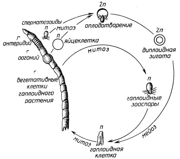
Fig. I.11. Reproduction cycle of Oedogonium
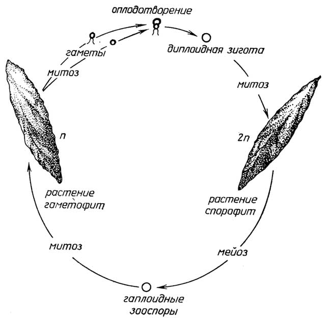
Fig. I.12. Reproduction cycle of Ulva
Chapter 4
PLANTS
These are multicellular organisms characterized by a photolithotrophic mode of nutrition, immotility, the presence of a cellulosic cell wall, the storage of polysaccharides in the form of starch, and the capacity for continuous growth throughout their entire lifespan.
§ 1.
Tissues of higher plants
Cellular differentiation in plants leads to the formation of several tissue types (Fig. I.13).
Meristematic tissue ensures the continuous growth of the plant. At the tips of roots and stems are cells of the apical meristem, which drives

Fig. I.13. Plant tissues: meristem (A), dermal (B), parenchyma (C), collenchyma (D), sclerenchyma (E), xylem tracheid cell (F), phloem sieve cell (tube) (G)
the apical growth of these organs. Growth in width of a trunk, stem, or root (secondary growth) is driven by the lateral meristem (the cambium layer). Meristematic cells are small in size, possess relatively large nuclei and thin walls, and lack vacuoles.
Dermal tissue protects the underlying tissues from damage and desiccation. It is represented by the epidermis of leaves and the cork layer of trunks, stems, and roots. Dermal tissue cells are characterized by thick walls; they produce hydrophobic substances that are either secreted to the exterior to coat the leaves (wax) or incorporated into the cell walls (suberin in cork).
Ground tissue constitutes the bulk of the plant. Its function is the synthesis and storage of nutrients. Ground tissue includes parenchyma.
Parenchyma consists of soft tissues found in leaves, bark, stems, roots, flowers, and fruits. Each parenchyma cell contains a large vacuole that may accumulate storage proteins, fats, tannins, and other substances. The vacuole frequently occupies the major volume of the cell. A specialized type of parenchyma is chlorenchyma (green parenchyma), the cells of which contain chloroplasts with chlorophyll and carry out photosynthesis.
Certain plant cells form part of specialized glands that produce resin, latex, mucilage, gums, and other substances. Sometimes these cells are classified as a special secretory tissue.
Cells of mechanical tissue perform a supporting function. Collenchyma and sclerenchyma are distinguished. Collenchyma is a resilient tissue found in leaf petioles, flowers, fruits, and stems. The primary difference between its cells and parenchyma cells is the presence of angular thickenings in the cell walls, which provides springiness and elasticity to plant organs, particularly stems. Sclerenchyma is a rigid plant tissue that provides mechanical strength to the stem and roots. Almost the entire space of sclerenchymal cells is occupied by a thickened, rigid cell wall. Sclerenchymal cells are dead. They frequently form long fibers that permeate the plant body. The shell of nuts and fruit pits is composed of such cells (stone cells).
Vascular tissue ensures the delivery of water and dissolved salts from the roots to all other cells, and synthesized nutrients to their storage sites. It also performs a supporting function. Vascular tissue is of two types: xylem (wood) and phloem (bast).
Xylem forms deep within the stem from long, thin cells—tracheids—which, by connecting end-to-end, transform into thin tubes reaching several meters in length, known as vessels. Following the death of the tracheids and the hardening of their cell walls (due to lignin deposition), bundles of tubes are formed within the plant through which water and dissolved salts are transported up the stem from the roots to the leaves.
Phloem also consists of sub-surface bundles of tubes formed, similarly to xylem, from cells joined end-to-end. Unlike xylem cells, however, phloem cells do not die, and the partitions between them do not disappear, instead bearing numerous pores. Through these phloem sieve tubes, nutrients produced in the leaves are transported to the stem and roots, while in the spring, the flow reverses from the roots and stem to the sprouting buds.
§ 2.
Plant organs
Leaf. The primary function of leaves is photosynthesis. The leaves of certain plants also perform the function of water storage (cacti) or nutrient storage (cabbage). The leaf surface consists of a protective tissue—the epidermis—beneath which lie the ground and vascular tissues.
Root. Plant roots ensure the absorption of water and dissolved salts from the soil. They also perform a supporting function, anchoring the plant in the soil. In some plants, roots that form root crops (carrots, beets) store nutrients. All types of tissues are present within the root.
The stem is the axial part of a plant shoot. Its main function is to support and optimally position the leaves relative to light. It also provides a connection between the roots and leaves, and in some plants, it serves as a storage site for nutrients (e.g., potato tubers). The stem structure contains all types of tissues, with vascular tissues being particularly well developed.
Flowers, cones, sporangia, antheridia, and archegonia are the reproductive organs of plants.
Flowers and cones are found in higher plants (such as apple trees and pines) that reproduce by seeds and exist independently solely as diploid sporophytes.
In lower plants, which can exist either as a haploid gametophyte or as a diploid sporophyte—either independently (ferns) or parasitizing the gametophyte (mosses)—we find: in gametophytes, antheridia, which produce spermatozoa, and archegonia, which produce egg cells; in sporophytes, sporangia, which produce asexual haploid spores that grow into gametophytes.
§ 3.
Specific Features of Plant Reproduction
Like protists, plants can reproduce both sexually and asexually.
Sexual reproduction is characteristic of all plants. It is associated with the formation of a diploid zygote as a result of fertilization—the first cell of a new organism (Figs. I.14–I.17). Higher plants, i.e., gymnosperms and angiosperms, represent the diploid sporophyte. In their reproductive organs—male cones (in gymnosperms, Fig. I.16) or flowers (angiosperms, Fig. I.17)—pollen is formed, which, via insects or wind, reaches other female cones or flowers, germinates, and reaches the egg cell located within the body of the sporophyte itself. Following fertilization, the seed gradually forms, inside which the diploid zygote develops into
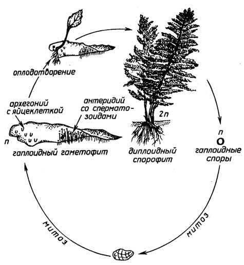
Fig. I.15. Fern reproduction cycle
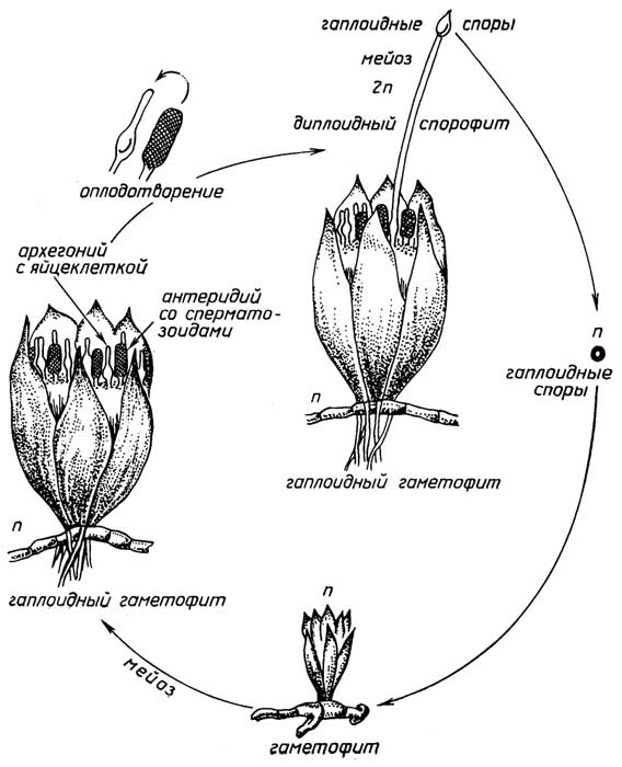
Fig. I.14. Moss reproduction cycle

Fig. I.16. Gymnosperm reproduction cycle
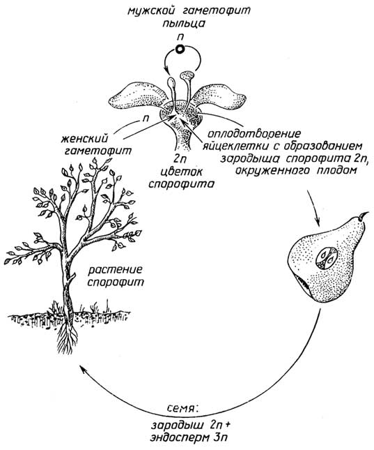
Fig. I.17. Angiosperm reproduction cycle
an embryo. Upon encountering favorable conditions, the seed grows into a new sporophyte.
Asexual reproduction in plants exists alongside sexual reproduction in two forms: spore formation and vegetative propagation.
Spore formation occurs in lower plants such as mosses (Fig. I.14), horsetails, and ferns (Fig. I.15). Unlike gymnosperms and angiosperms, the diploid sporophyte of such plants produces asexual haploid spores, from which haploid plants—gametophytes—grow under favorable conditions. Reproduction then proceeds sexually. The gametophyte produces gametes: spermatozoa and egg cells. An aqueous medium is required for fertilization because the motile spermatozoon must swim toward the egg cell located within the gametophyte body. A new sporophyte grows from the resulting zygote.
Vegetative propagation is found in both lower and higher plants. In some mosses and their variants, such as liverworts, the gametophyte is capable of reproducing by budding, when a part of its body in the form of a "brood cup" detaches and forms an independent plant. Vegetative propagation via a horizontal stem located on or beneath the soil surface, which generates new plants continuously, is found in both lower and higher plants. In mosses, such a horizontal "stem" is called a protonema. In higher plants, the aerial form of a creeping horizontal stem represents runners (as in strawberries), while the subterranean form is called a rhizome (potato tubers are thickened rhizomes). Vegetative propagation by cuttings and grafting is widely used in cultivating many crop plants.
§ 4.
Basic Principles of
Plant Classification
Because plant evolution did not proceed in a linear fashion, but rather resembles a branching tree diagram, it is difficult to arrange all plants in order of increasing structural complexity along a single line. The fundamental rule that allows plants to be grouped together is the commonality of their structure, development, and reproduction. The key criterion used to distinguish two groups of plants—one of which comprises the least developed forms—is the presence of vascular conductive tissues (Fig. I.18). In mosses, liverworts, and hornworts, belonging to the division Bryophyta, vascular tissues are absent. These small, simple plants are anchored in the soil by thin, root-like hair structures (rhizoids). Their stems are either absent or poorly developed. These

Fig. I.18. Principles of plant classification
plants share many features with multicellular algae.
The two distinguished plant groups also differ in that each predominantly exists in a haploid or diploid form. A double set of chromosomes promotes species survival; therefore, plants favoring the diploid form of existence have achieved better evolutionary development. In the less developed bryophytes, the gametophyte (the haploid form of existence) is better developed, upon which the sporophyte typically parasitizes, whereas in the more advanced vascular plants, the sporophyte is better developed.
Vascular plants, in turn, can be divided into two groups using an important evolutionary feature associated with an aromorphosis as a criterion: the presence or absence of an independently existing gametophyte generation. This distinction is linked to the transition from a mixed mode of reproduction (sexual—via spermatozoa and egg cells, and asexual—via spores) to a purely sexual pathway—via pollen and an egg cell located within the plant-sporophyte itself. Once this transition took place, the need for an aqueous medium for fertilization disappeared, meaning that plants became independent of abundant moisture and finally "conquered the land." Therefore, grouping ferns together with seed plants based solely on similarities in structure and the development of tissues and organs is not entirely justified due to the fundamental differences in the reproductive pathways of these two plant groups.
Chapter 5
ANIMALS. HUMANS
These are multicellular organisms with a chemoorganotrophic mode of nutrition, motility (in most species), the absence of a cellulosic cell wall, the storage of polysaccharides in the form of glycogen, and an inability to undergo continuous growth (increase in size) throughout their entire life span.
§ 1.
Tissues of Higher Animals
Cell differentiation in animals leads to the formation of tissues, which can be divided into several types based on their structural and functional characteristics, as well as their origin (see p. 77).
Epithelial tissueis formed by the cells of the outer layer of the skin (epidermis), as well as cells lining the inner surface of the digestive tract, respiratory and urogenital tracts, and exocrine and endocrine glands.
Functions of epithelial tissue: 1) protection against infection, as well as mechanical and chemical damage to underlying tissues; 2) absorption of water and nutrients; 3) secretion of various substances by glands.
Epithelial cells fit tightly together. Depending on their shape and functions, they are divided into several types, which can be reduced to two main ones: squamous and columnar epithelium (Fig. I.19 A, B). Epithelial tissue is evolutionarily older than all other tissues.
A special type of epithelial tissue is reproductive tissue. It consists of two categories of cells. The first category includes germ cells—the spermatozoon (Fig. I.19 C)
and the ovum (Fig. I.19 D), while the second includes the somatic cells of the testes and ovaries. Germ cells have a haploid set of chromosomes, whereas somatic cells have a diploid set. The process of sperm formation from spermatogonia (i.e., testicular cells) is called spermatogenesis. The transformation of oogonia (i.e., ovarian cells) into ova is called oogenesis. The transformation of spermatogonia and oogonia into germ cells (Fig. I.20)
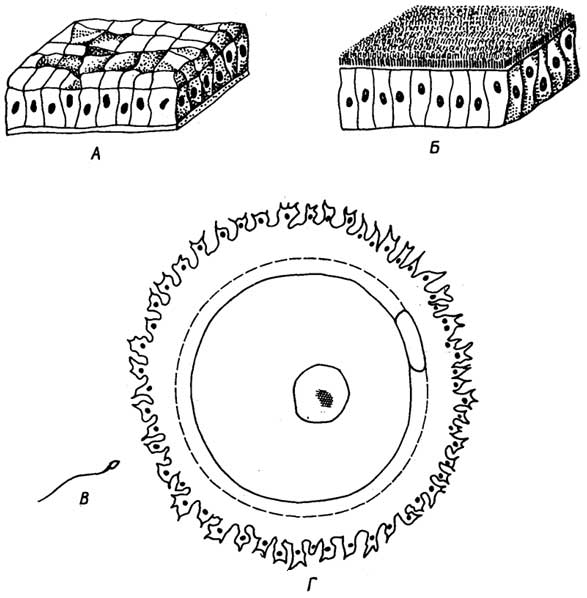
Fig. I.19.Main types of epithelial tissue: squamous (A) and columnar (B), as well as cells of a special type of epithelial tissue—reproductive tissue: spermatozoon (C) and ovum (D)
occurs via meiosis through the sequential formation of spermatocytes or oocytes, first primary, then secondary, and subsequently spermatids or ootids.
A significant difference between spermatogenesis and oogenesis is that primary and secondary spermatocytes divide to form identical cells, whereas primary and secondary oocytes divide to form different cells. The primary oocyte divides into two unequal parts: a larger secondary oocyte and a small first polar body (polocyte). During this division, the entire nutrient reserve located in the cytoplasm passes into the secondary oocyte, while the first polar body consists almost entirely of a nucleus. The secondary oocyte also divides into two unequal parts: a larger ootid and a small second polar body, which is also almost devoid of
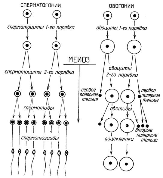
Fig. I.20.Spermatogenesis and oogenesis
cytoplasm; the first polar body may split into two identical second polar bodies during this time. All polar bodies soon degenerate and disappear.
Connective tissue is a system of cells that form the inner primary layer of the skin, tendons, ligaments, cartilage, bones, bone marrow, the subcutaneous fat layer and fat pads of organs, as well as fluid tissues such as blood and lymph.
Functions of connective tissue: 1) connecting—binds other tissues into a single whole and ensures metabolic exchange between them; 2) supporting—forms the skeleton, maintains the relative positioning of internal organs, and preserves body shape; 3) protective—protects underlying tissues and organs from hypothermia and mechanical damage; connective tissue cells seal wounds caused by mechanical trauma, prevent infection, foreign bodies, and toxins from entering the body, and remove them from it; 4) trophic—stores nutrients (fats).
A characteristic feature of connective tissue is that its cells are usually located at some distance from one another, embedded in a ground substance (Fig. I.21), although this does not prevent them from forming syncytia in certain cases. The ground substance, secreted by the cells, enables connective tissue to perform many of its functions.
Depending on the structure and composition of the ground substance and their functions, several types of connective tissue are distinguished: fibrous, cartilaginous, osseous, reticular, adipose, as well as blood and lymph.
Fibrous tissue is found throughout the body, connecting various tissues and organs. Examples include the inner layer of the skin (the one that remains after tanning and processing), as well as tendons and ligaments. The ground substance of fibrous tissue is a dense network of protein fibers running in various directions. Depending on the density of the ground substance and the number of cells per unit volume, it is subdivided into loose and dense (fibrous) connective tissue.
There are several cell types in connective tissue: fibroblasts, histiocytes, mast cells, and endothelial cells.
Fibroblasts are the primary cells of connective tissue; they form fibers and the intercellular ground substance, playing a major role in wound healing and scar formation.
Histiocytes are motile cells capable of engulfing and destroying foreign cells and particles, such as bacteria and viruses.
Mast cells are a special category of connective tissue cells present in many organs: the liver, spleen, lungs, blood vessel walls, brain, etc. Their unique feature is that they secrete physiologically active substances into the blood and tissue fluid, which influence, for example, blood clotting and pressure, as well as inflammatory, allergic, and immune responses.
Endothelial cells form a single-cell layer that constitutes the walls of blood and lymph capillaries; they also make up the inner lining of larger blood vessels and the heart.
Cartilage and bone tissues. The skeleton of very young vertebrate animals consists of elastic cartilage tissue, which is replaced with age by harder bone tissue, but in
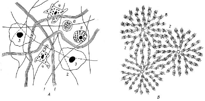
Fig. I.21.Connective tissue:
fibrous (A) and bone (B).
1 - protein fibers; 2 - ground substance; 3 - fibroblast; 4 - histiocyte; 5 - mast cell; 6 - lymphocyte; 7 - Haversian canal; 8 - osteoblast (bone cell)
certain lower vertebrates, such as sharks, the skeleton remains cartilaginous; in humans, cartilage tissue remains in the tip of the nose and the auricle, and forms the trachea, bronchi, larynx, etc. Bone tissue differs from cartilage in its greater rigidity (due to the deposition of insoluble calcium salts in the ground substance) and denser structure.
Reticular tissue forms the basis of the red bone marrow, spleen, lymph nodes, and certain structures such as the tonsils. Pluripotent stem cells of the red bone marrow play a primary role in hematopoiesis, giving rise to lymphoid and myeloid stem cells. The former are precursors to lymphocytes, while the latter are precursors to erythrocytes, megakaryocytes, macrophages, and granulocytes (see section "Blood").
Adipose tissue serves as both a protective cushion (subcutaneous fat layer, bed for internal organs) and a storehouse for high-calorie "fuel." Its cells (adipocytes) are almost entirely filled with fat. They form the basis of yellow bone marrow, which replaces red bone marrow in bone cavities with age.
Blood and lymph are specialized types of connective tissue. Their primary functions are binding (transport) and protection. Blood and lymph circulate throughout the body and, together with tissue fluid, play a crucial role in homeostasis—that is, maintaining the constancy of the internal environment of the body, specifically: the composition and concentration of substances, temperature, pH (acid-base balance), and ionic strength (water-salt balance). Blood transports oxygen (from the respiratory organs to all tissues), carbon dioxide (from tissues to the respiratory organs), nutrients (from the digestive organs to tissues in need), and metabolic waste products (to the excretory organs).
The protective function of blood and lymph lies in their ability to identify and neutralize foreign cells, viruses, harmful substances, and transformed autologous cells that have entered the body.
Blood consists of formed cellular elements (Fig. I.22)—erythrocytes, leukocytes, platelets—and an intercellular matrix called plasma.
Erythrocytes are red blood cells. In mammals, erythrocytes lose their nucleus during maturation. Erythrocytes of warm-blooded animals contain the iron-containing red pigment hemoglobin, which serves as a carrier of oxygen and carbon dioxide. They are produced by bone marrow cells and circulate in the blood for about 125 days, after which they are destroyed by leukocytes in the spleen and liver. 1 mm3 of blood in a healthy human contains 4–5 million erythrocytes.
Leukocytes are cells larger than erythrocytes; they possess a nucleus and are capable of proliferation, i.e., reproduction by mitosis. Due to their ability to significantly alter their shape and move in an amoeboid fashion, leukocytes can migrate against the blood flow, slipping between the cells of blood capillaries and tissues. The main function of leukocytes is phagocytosis—the destruction of infected, diseased, aging, transformed (tumor) cells, viruses, foreign macromolecular substances, and the production of protective proteins known as antibodies.

Fig. I.22. Formed elements of blood:
1 - erythrocytes; 2 - lymphocytes; 3 - monocytes; 4 - neutrophils; 5 - eosinophils; 6 - basophils; 7 - platelets
Based on their shape, structure, and functional features, leukocytes are conventionally divided into lymphocytes, monocytes (a type of macrophage), and granulocytes (or polymorphonuclear leukocytes).
Lymphocytes, in turn, are subdivided into several types: B, T, NK, L, K, and null cells. Granulocytes include neutrophils, eosinophils, and basophils. All leukocytes originate from red bone marrow cells. Most leukocytes are short-lived—ranging from a few hours to a few days—whereas certain types (immunological memory cells) persist in the body for up to ten years.
1 mm3 of blood in a healthy human contains 5–8 thousand leukocytes. The quantitative ratio of different leukocyte forms (the leukogram or leukocyte differential) is also relatively constant, changing in specific ways only during illnesses and various physiological states.
Platelets (blood platelets) are small (several times smaller than erythrocytes), anucleate cells derived from the cytoplasm of giant megakaryocytes in the red bone marrow. The function of platelets is to restore the integrity of damaged blood vessels and initiate hemostasis (blood clotting) at the site of injury. First, they "plug" the lesion with their bodies, and subsequently, a blood clot (thrombus) forms at the site. Platelets live for several days and are destroyed in the spleen and liver. 1 mm3 of blood in a healthy human contains 200–400 thousand platelets.
Blood plasma is a transparent yellowish fluid (containing 90% water) that remains after the blood cells are separated. Plasma proteins primarily perform protective and transport functions. Organic and mineral substances are also dissolved in the plasma. Plasma plays the leading role in both homeostasis and hemostasis.
Blood serum is a light yellow fluid that differs from plasma in that the protein fibrin has been removed. The function of blood fibrin is the formation of a blood clot (thrombus), which underlies the blood coagulation process. Serum forms spontaneously when a clotted blood clot contracts after some time and extrudes the serum.
Lymph is a colorless fluid found in lymphatic capillaries and vessels. It lacks erythrocytes but contains leukocytes. The composition of its liquid portion is similar to that of plasma. Like blood, when lymph escapes from vessels, it clots, albeit more slowly.
Functions of lymph: 1) facilitates substance exchange between tissues and blood; 2) destroys bacteria, viruses, tumor cells, and foreign macromolecular substances via leukocytes (phagocytosis); 3) ensures the absorption of lipids from the intestine and their transport into the blood.
Tissue fluid is an intermediate product in the conversion of blood—more precisely, plasma—into lymph. It is a component of the intercellular matrix of various tissues.
The blood of different individuals contains erythrocytes with a specific surface type, which depends on the presence of carbohydrate compounds (agglutinogens) of type A or B, or both A and B; in some individuals, agglutinogens are absent. Based on this, four blood groups are distinguished: I, II, III, IV, or 0, A, B, AB, respectively. The blood plasma of each group, except for the fourth, contains protein substances (agglutinins) capable of clumping and precipitating erythrocytes (causing agglutination) of other blood groups in accordance with Table I.1. For each race and nationality,
Table I.1
Blood groups and rules for blood transfusion
Blood group name |
Type of agglutinogens on the erythrocyte surface |
Type of agglutinins in blood plasma |
Recipient blood group |
Donor blood group |
I, or 0 |
— |
a and b |
I II III IV |
I |
a characteristic statistical ratio of blood groups is typical.
Human blood also varies due to the presence or absence of the Rhesus (Rh) factor (a carbohydrate compound on the erythrocyte surface), as well as other traits that play a significant role in massive blood transfusions or tissue transplantation.
Blood groups and the Rhesus factor must be taken into account when transferring blood from one person to another.
Muscle tissue is an aggregate of muscle cells. There are three types of muscle tissue (Fig. I.23): striated, smooth, and cardiac. Muscle cells are elongated in shape and form fibers (myofibrils). Striated muscle
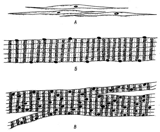
Fig. I.23. Muscle tissue: smooth muscle (A), skeletal muscle (B), and cardiac muscle (C)
muscle tissue includes skeletal muscles. The cardiac muscle (myocardium) also has striated fibers, but differs from skeletal muscles in the way muscle fibers are connected. The former are syncytia-like symplasts, while the latter form true syncytia. The structure of muscle tissue is determined by the contractile function of muscles.
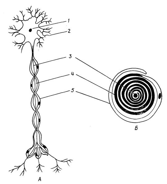
Fig. I.24. Nerve tissue cell — a neuron (A):
1 — neuron cell body; 2 — dendrites; 3 — axon; 4 — myelin sheath; 5 — neurilemma (Schwann cell). Formation of the axon myelin sheath by a neurilemma cell (B)
Nerve tissue is represented by cells from various parts of the nervous system: the brain and spinal cord, nerve ganglia, and nerve fibers. There are two main types of nerve cells: neurons and neuroglia.
Neurons (Fig. I.24) are the primary cells of nerve tissue, whose function is associated with the generation, reception, and transmission of signaling impulses. A structural feature of neurons is the presence of processes: longer ones (sometimes up to 1 m or more), called axons, and shorter ones, called dendrites. Axons and dendrites are outgrowths of the cell body and consist of cytoplasm covered, as usual, by a cell membrane. The axons and dendrites of the "white matter" neurons in the brain and spinal cord (i.e., the central nervous system), as well as the axons, dendrites, and cell bodies of neurons connecting the brain and spinal cord with various organs and tissues of the body (i.e., the peripheral nervous system), are additionally covered with a hydrophobic myelin sheath. Acting as a good insulator, it apparently facilitates better signal propagation.
Axons transmit an impulse from their neuron's body directly to an adjacent neuron or its dendrites, while dendrites receive the signal and convey it to their own neuron's body. There is no direct contact between the impulse-transmitting axon and the receiving neuron or its dendrite; they are separated by a small gap called a synapse. The transmission of an impulse across the synapse is mediated by specialized signaling substances called neurotransmitters, and occurs in only one direction — from the axon of one neuron to the body or dendrites of an adjacent neuron. The organs of the nervous system consist of numerous interconnected neurons, with each neuron linked simultaneously to hundreds, thousands, or tens of thousands of other neurons.
Neuroglia consists of supporting cells of nerve tissue. They fill the spaces between neurons, as well as between neurons and blood vessels of the brain, performing supportive, protective, and trophic functions. Specific neuroglial cells known as neurilemma cells, or Schwann cells (see Fig. I.24), synthesize the myelin sheath for axons, dendrites, and the surface of certain neurons.
§ 2.
Organs of Higher Animals
and Humans
Animal organs are more numerous than those of plants. They are typically organized into systems. Higher animals and humans possess 11 organ systems.
The Circulatory System
The circulatory (cardiovascular) system performs a connective (transport) and protective function. It ensures the transport of oxygen and metabolic exchange between organs, delivering nutrition and chemical energy while removing metabolic waste products. The cardiovascular system also mediates the protective functions of blood and lymph. It comprises the circulatory and lymphatic systems, including the heart, blood and lymphatic vessels, blood, lymph, and tissue fluid. It can also include hematopoietic organs — the red bone marrow, spleen, liver (which is also part of the digestive system), and the thymus, an endocrine organ that plays a crucial role in the protective function of blood (p. 68).
The heart (Fig. I.25) is a muscular pump that drives blood through the vessels. In addition to the contractile myocardium (cardiac muscle tissue), it contains specialized muscle tissue that retains embryonic properties and forms the heart's conduction system, which generates electrical impulses responsible for rhythmic heart contractions.
The heart is enclosed in a fluid-filled connective tissue sac called the pericardium. The inner surface of the pericardium and the outer surface of the myocardium are lined with smooth endothelial cells to reduce friction during contractions.
Based on their functions and wall structure, blood vessels are divided into arteries, veins, and capillaries. Arteries carry blood away from the heart to organs and tissues. Emerging from the heart are the pulmonary artery, which directs blood to the lungs, and the aorta (the main and largest artery), which branches into other arteries shortly after leaving the heart. Arteries, in turn, branch into smaller vessels called arterioles and then into a vast network of capillaries. The walls of arteries and arterioles consist of three layers (see Fig. I.25): an outer durable and elastic connective tissue layer, a middle smooth muscle layer, and an inner connective tissue layer lined internally with a layer of smooth, flat endothelial cells. In most arteries, the connective tissue of the inner layer forms a strong yet elastic membrane.
Veins transport blood from organs and tissues back to the heart. Within organs and tissues, blood passes from the capillaries of the arterial system into venous capillaries, which converge into larger venules and subsequently into larger veins. Blood returns to the heart via the superior (from the lungs) and inferior (from other organs) vena cava. Thus, the arterial and venous beds are connected via a capillary network. Metabolic exchange between blood and tissue fluid occurs across the capillary walls, which consist solely of endothelial cells. The walls of veins and venules share the same three layers as arteries and arterioles, but are noticeably thinner. In most veins, the connective tissue of the inner layer lacks an elastic membrane. The main distinguishing feature of veins is the presence of valves situated at regular intervals, which allow blood to flow in only one direction. A special role in the circulatory system is played by the hepatic portal vein, which channels blood from the spleen and digestive organs (stomach, pancreas, intestines) into the liver.

Fig. I.25. Heart (A):
1 — right atrium; 2 — left atrium; 3 — right ventricle; 4 — left ventricle; 5 — inferior vena cava; 6 — superior vena cava; 7 — pulmonary vein; 8 — pulmonary artery; 9 — aorta. Structure of arteries (B):
1 — outer connective tissue layer; 2 — middle muscle layer; 3 — inner connective tissue layer; 4 — endothelial lining of the inner layer; 5 — lumen of the artery

Fig. I.26. Respiratory system (A):
1 — trachea; 2 — bronchi; 3 — bronchioles; 4 — lung. Branching of bronchioles (B):
1 — bronchioles; 2 — alveolar ducts; 3 — alveolar sacs; 4 — alveoli
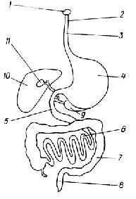
Fig. I.27. Digestive system:
1 — mouth; 2 — pharynx; 3 — esophagus; 4 — stomach; 5 — duodenum; 6 — small intestine; 7 — large intestine; 8 — rectum; 9 — pancreas; 10 — liver; 11 — gallbladder
From there, blood is channeled via the hepatic vein and subsequently the inferior vena cava into the heart.
Lymphatic vessels include both small capillaries and larger vessels. The walls of lymphatic capillaries also consist of endothelial cells and are permeable to substances dissolved in tissue fluid. A fundamental difference between lymphatic and blood capillaries is that lymphatic capillaries are blind-ended. Like blood capillaries, they absorb substances from the tissue fluid. Subsequently, lymphatic capillaries merge into larger vessels, which in turn combine into even larger ones. The largest of these, the thoracic duct, empties into the left subclavian vein, thereby connecting the lymphatic network with the circulatory system.
Thus, fluid and all dissolved substances return from the tissues to the blood via both blood and lymphatic capillaries. Large lymphatic vessels, much like veins, feature valves at regular intervals that permit lymph to flow in only one direction. At the junctions of lymphatic vessels lie lymph nodes — specialized filters consisting of dense cellular tissue with numerous narrow and tortuous pathways. Within lymph nodes, lymphocytes proliferate, antibodies are produced, and bacteria, transformed (cancerous) cells, and other foreign bodies are trapped and destroyed.
Hematopoietic organs continuously replenish the blood. The red bone marrow, located within bone cavities, produces formed elements of the blood: erythrocytes, leukocytes, and platelets. The spleen acts as a blood reservoir; additionally, specialized cells within it phagocytize "old" erythrocytes and platelets. The liver serves as an even larger blood depot, capable of storing up to 20% of the total blood volume, which can be mobilized into circulation when needed. Much like the spleen, the liver performs a cleansing function (via Kupffer cells) and supplies the blood with numerous vital components.
The Respiratory System
The respiratory system supplies the body with oxygen and rids it of carbon dioxide.
It consists of the nasal cavity, larynx, and trachea, which branches into the right and left bronchi, which further divide into bronchioles. The latter terminate in several alveolar sacs, whose walls feature pouch-like protrusions called alveoli (Fig. I.26).
The walls of the bronchi and trachea consist of three layers: an inner mucosal epithelial layer, a middle smooth muscle and cartilaginous ring layer, and an outer connective tissue layer. The epithelial cells bear cilia, whose beating sweeps bacteria inhaled with air, as well as dust particles and other debris, back up into the larynx. As the bronchi branch further, their outer and middle wall layers disappear, and the ciliated epithelium is replaced by standard squamous epithelium.
Gas exchange in the lungs between the air and blood occurs via diffusion through the walls of the alveoli, which consist solely of a layer of epithelial cells, and the endothelium of the blood capillaries densely blanketing the alveoli. The elasticity of the lungs is ensured by an intervening elastic fibrous connective tissue that separates and supports the alveoli. The lungs are also permeated by numerous nerves, including both sensory nerves and those regulating the inhalation-exhalation process.
Digestive System
The digestive system is designed to extract the substances and energy necessary for the organism from food products. It consists of (Fig. I.27) the oral cavity, pharynx, esophagus, stomach, intestines (duodenum, small, large, and rectum), liver with the gall bladder, and pancreas.
In the oral cavity, food is preliminarily ground down and moistened with saliva secreted by the salivary glands. Saliva contains certain enzymes that partially break down carbohydrates, as well as mucus, which makes the food bolus easy to swallow. Through the pharynx and esophagus, food enters the stomach. The walls of the stomach are quite thick, consisting of three layers: an inner folded mucous layer represented by epithelial cells, a middle muscular layer, and an outer layer of fibrous connective tissue. The cells of the mucosal epithelium secrete an acidic (pH 1.5–2) enzyme-rich gastric juice, which initiates protein digestion. The contraction and relaxation of the smooth muscles in the middle layer of the stomach walls ensure the movement and mixing of the food chyme. The outer surface of all sections of the digestive tract is additionally covered by a tough, slippery serous membrane—the peritoneum. The walls of the stomach are permeated with nerves, blood vessels, and lymphatic vessels. A similar three-layered structure is found in the walls of the intestines, which begin with the relatively short (about 25 cm) duodenum. Here, through ducts, bile from the liver and pancreatic juice from the pancreas are emptied. The mucosal epithelial cells of the duodenum themselves secrete intestinal juice. Through the combined action of all three components, which have a pH of 7–8, the final breakdown of proteins, fats, and carbohydrates occurs in the duodenum and small intestine. The absorption of digestion products also takes place in the small intestine. The products of protein and carbohydrate digestion are absorbed into the blood capillaries of the intestinal walls and are subsequently transported via the portal vein to the liver.
The products of fat digestion are absorbed by the mucosal epithelial cells of the small intestine, within which fats are resynthesized from these products. The latter are absorbed by lymphatic capillaries and then carried via lymphatic vessels into the blood of the left subclavian vein. Water is absorbed in the large intestine. This region also harbors a multitude of beneficial microbes that process food residues—such as breaking down cellulose—and synthesize certain substances essential to the body. The contents of the large intestine gradually transition into a semi-solid state and are propelled into the rectum, from which they are eliminated as feces. Feces represent undigested food residues, unabsorbed or unnecessary substances, as well as a large quantity of bacteria and their metabolic byproducts. Beyond synthesizing bile—which is concentrated in the gall bladder—the liver carries out numerous conversions of some substances into others, thereby ensuring the constancy of chemical composition and their ratio across various tissues and organs.
Excretory System
The excretory system ensures the removal of metabolic waste products from the organism.
It primarily comprises the kidneys and urinary tract, which eliminate the bulk of "waste." The organs of the respiratory system (excreting carbon dioxide), digestive system (eliminating "waste" with feces), and integumentary system (the skin excretes 5–10% of metabolic waste via sweat, largely the same substances found in urine) also possess excretory functions. In addition to its excretory function, the kidneys play a crucial role in homeostasis—that is, maintaining the constancy of the internal environment of the body, including the blood.
The kidneys are paired organs (Fig. I.28). Each kidney consists of a large number of functional units called nephrons, whose structure is illustrated in the figure. The renal artery branches within the kidney into arterioles, which in turn branch inside the nephron into a spherical cluster of capillaries known as the renal glomerulus. The afferent arteriole enters the glomerulus, while the efferent arteriole exits it. The renal glomerulus is housed within a double-walled cup called Bowman's capsule, from which extends a tubular vessel referred to as the renal tubule. The walls of the renal tubule consist of a single layer of epithelial cells. Upon emerging from the renal glomerulus, the efferent artery branches extensively into capillaries that densely and intimately envelop the beginning of the renal tubule (the proximal tubule) and its end (the distal tubule). The proximal and distal ends of the renal tubule, intertwined with numerous capillaries, together with the renal glomerulus enclosed in Bowman's capsule, constitute the portion of the nephron located in the renal cortex.

Fig. I.28. Excretory system.
Kidneys (A): 1 - left kidney; 2 - cortex of the right kidney; 3 - medulla of the right kidney; 4 - renal pelvis; 5 - ureters; 6 - urinary bladder; 7 - urethra.
Nephron (B): 1 - renal glomerulus; 2 - Bowman's capsule; 3 - afferent arteriole; 4 - efferent arteriole; 5 - renal tubules; 6 - loop of Henle; 7 - capillary network; 8 - venule; 9 - collecting duct; 10 - contribution from other nephrons
The middle section of the renal tubule, known as the loop of Henle, is located in the renal medulla in the majority of nephrons and is likewise enveloped by numerous capillaries that converge to form a venule. These venules subsequently merge into the renal vein. The distal part of the renal tubule transitions into the collecting duct, through which urine enters a small cavity within the kidney called the renal pelvis. From the renal pelvis, urine travels via the ureter into the urinary bladder, from which it is expelled from the body through the urethra.
Urine formation in each nephron begins with the filtration of blood in the renal glomerulus through the inner wall of Bowman's capsule into the proximal section of the renal tubule. Filtration is driven by the blood pressure gradient between the entrance and exit of the renal glomerulus, which arises because the efferent arteriole has a smaller diameter than the afferent one. Only low-molecular-weight substances, salts, and water pass from the blood into the resulting filtrate.
The conversion of filtrate into urine occurs within the renal tubules, firstly through the reabsorption of essential substances (glucose, amino acids, water, etc.) back into the blood capillaries surrounding the renal tubules, and secondly through the secretion of additional low-molecular-weight "wastes" from the blood capillaries into the renal tubules. Unlike filtration, reabsorption and secretion occur against a concentration gradient and therefore require energy expenditure. Transport processes that consume energy, as in this case, are referred to as active transport. Precise measurements demonstrate that the work performed by a kidney (per unit of its mass) even exceeds the work performed by the heart muscle. Through these active processes of reabsorption and secretion, the kidneys play a role in homeostasis that is no less significant than that of the liver.
Nervous System
The nervous system regulates and coordinates the functioning of all other body systems during interaction with the external environment. It consists of the brain and spinal cord, nerve nodes (ganglia), and nerves.
Structurally, the nervous system is subdivided into the central nervous system, which includes the brain and spinal cord, and the peripheral nervous system, which consists of ganglia and bundles of nerve fibers (nerves) connecting the brain and spinal cord to all other organs and tissues of the body.
Functionally, the nervous system is divided into the somatic nervous system, which controls the contraction and relaxation of skeletal muscles and bodily movement, and the autonomic (or vegetative) nervous system, which controls the activity of smooth muscles, the heart, and other internal organs.
Central Nervous System
Represented by numerous divisions.
Brain
The brain is a highly complex structure (Fig. I.29). Its bulk is occupied by the cerebral hemispheres—a paired structure consisting of gray matter and white matter. Gray matter
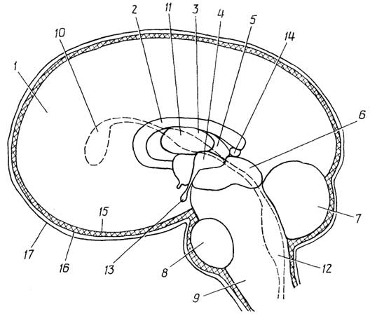
Fig. I.29. Brain:
1 - cerebral hemispheres; 2 - corpus callosum; 3 - thalamus; 4 - hypothalamus; 5 - epithalamus; 6 - midbrain; 7 - cerebellum; 8 - pons; 9 - medulla oblongata; 10 - first and second cerebral ventricles; 11 - third cerebral ventricle; 12 - fourth cerebral ventricle; 13 - pituitary gland; 14 - pineal gland ( epiphysis ); 15, 16, 17 - pia mater, arachnoid mater, and dura mater, respectively
of the brain represents an accumulation of neuronal cell bodies and their unmyelinated processes. In higher animals and humans, the gray matter of the brain forms a thin, heavily folded layer known as the cerebral cortex. The most complex forms of nervous activity are associated with its function: learning, memory, perceptual analysis, and creativity—in other words, mental activity. Beneath the cortex lies the white matter of the brain, composed primarily of myelinated neuronal processes that connect the cerebral cortex with other regions of the brain. Discrete masses of gray matter located within the cerebral hemispheres form subcortical nerve nodes, or basal ganglia, also referred to as nerve centers, which serve as intermediate relay stations for collecting, preprocessing, and transmitting nerve impulses further to other nerve centers, to the cerebral cortex, or, conversely, from the cortex to the target organs. The cerebral hemispheres together with the basal ganglia are called the telencephalon.
The brainstem (basal part of the brain) connects the telencephalon to the spinal cord. It is responsible for the reflex control of emotions, instincts, and muscle movement, and consists of the diencephalon, midbrain, medulla oblongata, and hindbrain. It houses the nerve centers of the autonomic nervous system.
The diencephalon contains various nerve centers, including those of the autonomic nervous system. The largest nerve centers of the diencephalon are the thalamus, which integrates all types of sensory impulses (thermal, pain, etc.) entering the brain, and the hypothalamus, which acts as the connecting link between the nervous and endocrine systems. Two important endocrine glands are also located here: the pituitary gland and the pineal gland (epiphysis). The hypothalamus regulates the overall level of excitability of the central nervous system. It plays a vital role in body temperature regulation. Because the hypothalamus is closely linked to one of the most important endocrine glands—the pituitary gland—the regulatory influence of the nervous system over many metabolic processes and the activity of the endocrine, cardiovascular, digestive, and excretory systems is executed precisely through the pituitary gland.
The midbrain connects the diencephalon with the medulla oblongata. It contains neural centers that regulate the activity of certain muscles, such as those of the eyes and ears in animals, as well as skeletal muscle tone.
The medulla oblongata houses the autonomic nervous system's neural centers, which control the functioning of numerous internal organs (the heart, lungs, digestive system, etc.). Additionally, it contains neural centers that process impulses from taste and auditory receptors, as well as organs of equilibrium.
The hindbrain comprises the cerebellum and the pons. The cerebellum is located in the posterior occipital region of the brain, behind the medulla oblongata and partially behind the midbrain. It consists of two hemispheres interconnected by the pons, which arches across the junction of the medulla oblongata and the midbrain. The cerebellar cortex features numerous folds. The neural centers of the cerebellum ensure the coordination of various muscle actions.
Spinal Cord
The spinal cord (Fig. I.30) lies within the vertebral canal and has a tubular shape. It plays a primary role in mediating most reflexes and participates in transmitting impulses between organs and tissues and the brain, and vice versa. The spinal cord consists of an inner gray matter, which appears butterfly-shaped in cross-section and represents an accumulation of neuronal cell bodies. Externally, it is surrounded by white matter, composed mainly of bundles of myelinated axons and dendrites.
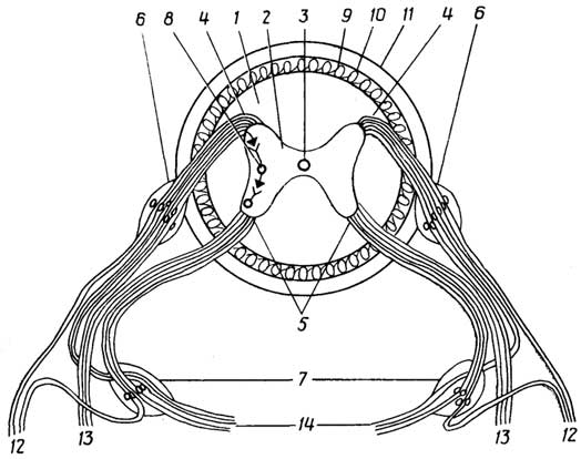
Fig. I.30. Spinal cord:
1 - white matter; 2 - gray matter; 3 - central canal; 4 - dorsal roots; 5 - ventral roots; 6 - dorsal root ganglia with sensory neurons; 7 - sympathetic nervous system ganglia with motor neurons; 8 - interneuron of the reflex arc; 9, 10, 11 - pia mater, arachnoid mater, and dura mater; 12, 13 - dorsal and ventral branches innervating the skin and muscles of the posterior and anterior parts of the body, respectively; 14 - autonomic branches innervating internal organs
Structural similarities between the brain and spinal cord. Running longitudinally through the center of the spinal cord is the central canal, filled with cerebrospinal fluid, which is compositionally similar to blood plasma. As this canal enters the brainstem, it expands in four regions. The four cavities formed by these expansions are called cerebral ventricles. The first and second are located in the cerebral hemispheres, the third in the diencephalon, and the fourth in the medulla oblongata. They are densely entwined with blood capillaries. Both the spinal cord and the brain are enveloped in three meninges: the inner pia mater (composed of neuroglial cells), the middle arachnoid mater (made of loose connective tissue), and the outer, relatively tough dura mater (composed of dense connective tissue). The space between these membranes is filled with cerebrospinal fluid.
Peripheral Nervous System
The peripheral nervous system consists of nerves connecting the brain and spinal cord to all organs and tissues. They are primarily bundles of axons and dendrites. Nerve plexuses are networks formed by the convergence of such bundles. The cell bodies of peripheral neurons reside either within the central nervous system or outside it, forming structures known as ganglia. Nerve impulses enter the brain or spinal cord via sensory (afferent) neurons and exit via motor (efferent) neurons. Communication between sensory and motor neurons is mediated by interneurons. Together, they form a functional unit—the reflex arc. Sensory fibers enter the spinal cord through dorsal roots, while motor fibers exit through ventral roots (see Fig. I.30). The dorsal and ventral roots soon merge into a single nerve trunk, which then divides again into three branches. The first two innervate the skin and muscles, while the third supplies the internal organs and thus belongs to the autonomic nervous system.
Autonomic Nervous System
Unlike the somatic nervous system, the autonomic nervous system is involuntary, meaning it is not subject to conscious control. It is subdivided into sympathetic and parasympathetic divisions. Most internal organs receive dual innervation from both the sympathetic and parasympathetic systems, though certain organs are innervated by only one. The two systems exert opposing effects on organs with dual innervation: while sympathetic impulses typically "accelerate" organ activity, parasympathetic impulses "inhibit" it, and vice versa. The normal functioning of internal organs depends heavily on the coordinated action of both the sympathetic and parasympathetic systems.
Structurally and functionally, the nervous system comprises the pyramidal tract and extrapyramidal system, sensory structures, the limbic system, and the reticular formation.
The pyramidal tract (system) is a collection of neural centers and efferent nerve pathways in the brain and spinal cord through which voluntary movement commands are transmitted from the cerebral cortex, via spinal cord neurons, to the motor nerves emerging from the spinal cord within the ventral roots.
The extrapyramidal system is a network of subcortical neural centers within the cerebral hemispheres and brainstem that participate in movement regulation, muscle tone control, and emotional expressions such as laughing or crying.
Sensory structures include nerve endings, afferent fibers, sensory neurons, and specialized projection areas across various regions of the central nervous system responsible for the perception, transmission, and processing of signals originating from receptors in virtually all body tissues and organs.
The limbic system is a complex of brain structures located in the cortex, subcortex, and deep within the cerebral hemispheres, which governs instinctive behavior and emotional states. It also plays a role in autonomic regulation and the formation of certain types of memory.
The reticular formation is a network of central brain structures involved in regulating the overall excitability level of the central nervous system, particularly the cerebral cortex. Specifically, the reticular formation modulates the sleep-wake cycle.
Endocrine and Exocrine Gland Systems
Endocrine glands are ductless glands that secrete various chemical messengers called hormones directly into the bloodstream. Even in extremely low concentrations, these substances precisely target and regulate the activity of other organs and tissues. Alongside the nervous system, the endocrine system plays a vital role in coordinating the functional activity of all body systems. Unlike neural regulation, endocrine glands provide humoral regulation. The activity of the endocrine glands themselves is regulated both neurally and humorally—meaning hormones secreted by certain glands influence the activity of others, operating via feedback loops commonly found in living organisms. Furthermore, different glands often secrete overlapping sets of hormones, so the functions of one gland can occasionally be partially assumed by another. Thus, the functioning of the endocrine glands is deeply interconnected. The endocrine system comprises (Fig. I.31): the pituitary gland, pineal gland, thyroid and parathyroid glands, thymus, adrenal glands, pancreas, gonads, as well as endocrine cells scattered in the mucosa of the digestive tract and heart.
The pituitary gland is located at the base of the diencephalon, directly beneath the hypothalamus. It consists of an anterior and a posterior lobe, which have distinct embryological origins. Hormones of the anterior lobe (adenohypophysis) regulate the function of numerous other endocrine glands, including the thyroid,
|

the adrenal glands, the endocrine cells of the pancreas (islets of Langerhans), the testes, and ovaries, as well as exocrine glands such as the mammary glands. Hormones of the posterior lobe (neurohypophysis) stimulate the contraction of smooth muscle and mammary glands, induce vasoconstriction of blood capillaries, and enhance water reabsorption by blood capillaries from the renal tubules.
The pineal gland is situated in the diencephalon. Its hormones exert an inhibitory effect on the function of the gonads and the adrenal cortex.
The thyroid gland is located in the neck region. Its hormones regulate the rate of metabolic reactions throughout the body.
The parathyroid glands (four in total) are situated on the posterior surface of the thyroid gland, occasionally embedded within its tissue. The hormones they secrete regulate calcium and phosphorus metabolism.
The thymus (or thymus gland) consists of two lobes flanking the trachea on the right and left. It is well-developed in newborns and grows until puberty, after which it undergoes involution, which in some cases leads to atrophy. The thymus plays a crucial role in immune system development, and the maturation of specific types of lymphocytes depends on its function. Thymic hormones significantly influence organismal development.
The adrenal glands are paired organs, with each located atop a kidney. They consist of two independent parts: an inner dark-brown medulla and an outer light yellowish-pink cortex. The adrenal medulla is composed of chromaffin cells, which represent a specialized type of nervous tissue. Its hormonal activity is regulated by the sympathetic nervous system. The hormones of the adrenal medulla (adrenaline and noradrenaline) play a vital role in mobilizing the body during stress—under peak physical and mental strain, in moments of danger, anxiety, or emergency circumstances. The adrenal cortex secretes various steroid hormones that influence many metabolic processes, the body's water-salt balance, and the lymphocyte count in the blood. The organism's resistance to stress and infections, as well as the adequacy of inflammatory and allergic responses, depends on the normal functioning of the adrenal cortex.
The pancreas consists of two types of cells: endocrine and exocrine. Endocrine cells are arranged within the pancreatic tissue in clusters known as the islets of Langerhans. They secrete various hormones that primarily regulate carbohydrate metabolism and, to some extent, lipid metabolism.
The gonads—testes (in males) and ovaries (in females)—secrete male and female sex hormones into the bloodstream, with the former predominating in males and the latter in females. This ratio determines the physical and behavioral traits characteristic of each sex.
The temporary glands in females include the corpus luteum and the placenta, whose hormones ensure the normal progression of pregnancy.
Cells of the gastric and intestinal mucosa also secrete several hormones into the bloodstream that regulate local digestion (at the level of a specific segment).
In addition to its excretory function, the kidneys act as an endocrine gland by releasing hormones into the blood that regulate erythropoiesis (the maturation of red blood cells).
Recent studies have shown that the heart functions as an important endocrine gland, releasing into the bloodstream a hormone involved in the regulation of metabolism, blood pressure, and water-salt balance.
Exocrine glands are glands that secrete their products not into the blood, but through ducts either to the exterior or into other internal organs. These include the skin's sweat and sebaceous glands, as well as salivary and mammary glands. Exocrine pancreatic cells, known as acinar cells, produce digestive enzymes that enter the intestine via pancreatic juice. The liver, acting as an exocrine gland, secretes bile, which flows through ducts into the duodenum. The prostate gland, seminal vesicles, and Cowper's glands are found exclusively in males and are located beneath the urinary bladder. The secretion they release into the genital ducts ensures the viability of spermatozoa within the seminal fluid.
Integumentary System
Represented by the skin, which serves as the body's outer shell. Its primary purpose is to protect the organism from external environmental factors while maintaining internal homeostasis. In other words, the skin protects against minor mechanical and chemical insults, against the harmful effects of

Fig. I.32. Skin:
1 - epidermis; 2 - upper layer of the dermis composed of dense fibrous connective tissue; 3 - lower layer of the dermis composed of loose connective tissue; 4 - adipose tissue; 5 - muscular tissue; 6 - hair; 7 - arrector pili muscle; 8 - sebaceous gland; 9 - sweat gland; 10 - nerve endings of the hair follicle; 11, 12, 13 - tactile receptors of varying sensitivity; 14 - free nerve endings (nociceptors); 15, 16 - heat and cold receptors, respectively
ultraviolet radiation, microbial penetration, overheating, and heat loss. The exocrine glands of the skin—sweat and sebaceous glands, as well as mammary glands, which originate from the same tissue as the skin—are organs of the excretory system. The skin consists (Fig. I.32) of a very thin outer layer, the epidermis, devoid of blood capillaries, and a thicker underlying layer, the dermis, which is permeated by numerous blood and lymphatic capillaries as well as nerve fibers. The epidermis is composed of epithelial cells, while the dermis consists of fibrous connective tissue cells and fat cells. The upper layer of the dermis is a very dense network of protein fibers formed by connective tissue cells called fibroblasts. This layer provides the mechanical strength and elasticity of the skin.
Skeletal System
Bones and their junctions together form the skeleton—a container for vital organs and a repository of mineral compounds. The skeleton performs a supportive as well as an architectural function, shaping the body. The rigid bones of the skeleton also protect internal organs. The human skeleton consists of more than 200 bones, as well as numerous cartilages, ligaments, and tendons. In humans and other advanced animals, the skeleton is located inside the body, whereas in other, more primitive animals (crayfish, crabs, snails, and certain insects), the skeleton is an external structure.
Cartilage is a semi-rigid connective tissue structure. It is found wherever resilient, dense tissue is required: the articular surfaces of bones, the larynx, trachea, bronchi, semi-rigid connections between the ribs and the sternum, etc. The auricles of the ears and the tip of the nose are also constructed from cartilage.
Ligaments are elastic connective tissue structures that connect bones to one another.
A joint is the site of a movable connection between two bones, typically enclosed in an articular capsule formed by ligaments. Its inner portion, the joint cavity, is filled with synovial fluid, which is compositionally similar to lymph but also contains mucous substances that act as a lubricant.
Tendons are non-stretchable, semi-rigid connective tissue structures capable of withstanding high tensile loads. They connect muscles to bones.
Muscular System
The muscular system, represented by numerous body muscles, together with the skeleton, shapes the body and ensures both the movement of its individual parts and locomotion of the entire organism.
Muscles in humans and mammals are divided into three types: skeletal, cardiac, and smooth. Skeletal muscles provide movement for the body or its parts; cardiac muscle (myocardium) drives the heart as a pumping engine; smooth muscles, which form the basis of the walls of internal organs and blood vessels, maintain vascular tone and lumen, as well as those of hollow organs.
Muscle activity is regulated via neurohumoral pathways—that is, through the nervous system and physiologically active substances, including hormones. Voluntary control is exclusive to skeletal muscles.
Sensory Organs
It is generally accepted that higher animals and humans possess five types of senses: sight, hearing, touch, smell, and taste, through which the external environment is perceived. For the organism's vital activity, signals originating from the internal environment (perception of internal state) are just as important as information from the outside world, even though the sensations arising from them are difficult to precisely characterize and define. Animals and humans also possess a sense of balance (graviception), which relies on the functioning of both a dedicated organ and other sensory organs, including those that perceive the internal state of the body.
The functioning of all types of sense organs is based on the ability of their receptor cells to be stimulated and subsequently transmit the generated nerve impulse to the central nervous system.
The organ of vision is the eye. Its receptor cells, cones and rods, react to light. The former are responsible for vision in bright light, as well as the perception of color, shape, and details of objects, while the latter are responsible for dim-light vision and night vision.
The organ of hearing consists of the outer, middle, and inner ear. Inside the inner ear are auditory receptors—hair cells—which perceive sound signals.
The organs of touch are located in the inner layer of the skin (see Fig. I.32). They are various types of nerve endings extending from sensory neurons: either freely branching (responsible for pain sensations) or specialized structures, such as capsules (responsible for tactile sensations, as well as the perception of cold and warmth). It is believed that exceeding a certain threshold of stimulation in tactile and temperature receptors also triggers pain sensations.
The organs of smell. Olfactory receptor cells are situated among the epithelial cells of the upper nasal passages, where air reaches exclusively via diffusion.
The organs of taste. Taste receptor cells are located within taste buds, which are found mainly on the tongue and partially on the soft palate, the posterior wall of the pharynx, the tonsils, the epiglottis, and, in early childhood, on the lips as well.
The organs of balance (gravity) comprise the vestibular apparatus, anatomically situated in the inner ear, alongside numerous kinesthetic receptors (proprioceptors) located in muscles, ligaments, joint capsules, and connective tissue structures, which respond to stretch and compression. The sense of balance is also supported by the organs of vision and tactile receptors in the skin.
Receptors for environmental perception organs (vision, hearing, touch, smell, and taste) are called exteroceptors. Receptors that perceive the internal state of the organism are called interoceptors, or receptors of visceral sensitivity. They are located in internal organs and blood vessels. Feelings of thirst, satiety, nausea, vomiting, dizziness, and the urge to urinate or defecate are wholly or partially linked to their signals. This group also includes the receptor cells of the vestibular apparatus and proprioceptors. Stimulation of sense organs occurs via mechanical means (tactile, auditory, vestibular, proprioceptive, and partly interoceptive), chemical means (olfactory, gustatory, pain, and some interoceptive), or physicochemical/photochemical means (visual).
Reproductive organs
Reproductive (sex) organs ensure the generation of offspring. They are divided into internal and copulatory organs.
The internal reproductive organs in males are the testes and accessory sex glands: the prostate, Cowper's glands, and seminal vesicles. In addition to synthesizing sex hormones, the testes produce male germ cells—spermatozoa—while the accessory sex glands produce fluid that ensures sperm viability.
The internal reproductive organs in females are the ovaries, oviducts (fallopian tubes), uterus, and placenta. Besides sex hormones, the ovaries produce ova (egg cells), which are fertilized by spermatozoa in the fallopian tube. The uterus is where embryonic development and fetal gestation take place. The placenta is a temporary organ that forms during pregnancy from the inner lining of the uterus. It is a spongy membrane that separates the fetal blood from the maternal blood, yet allows the fetus to receive nutrients and oxygen from the maternal organism while eliminating carbon dioxide and metabolic waste. The placenta also functions as an endocrine gland, producing hormones that regulate the course of pregnancy.
Copulatory (external) reproductive organs facilitate the meeting of spermatozoa and the ovum.
Riculoendothelial (macrophagic)
system
Unlike other organ systems in humans and higher animals, the reticuloendothelial system is not a collection of organs—often built from multiple types of tissue and united by one or more shared functions—but rather a collection of cells with identical functions distributed across different tissues and organs. The primary function of reticuloendothelial cells is to protect the organism against bacteria, viruses, and foreign particles that have entered the body or formed within it as a result of microbial activity or metabolic disruption. This defense is carried out through phagocytosis.
The cells of the reticuloendothelial system include: blood monocytes and granulocytes (neutrophils), as well as macrophagic cells in various tissues and organs—such as histiocytes in connective tissue, osteoclasts in bone tissue, and Kupffer cells in the liver. In other organs and tissues (spleen, lungs, bone marrow, lymph nodes), macrophages lack specialized names. Neuroglial cells also possess phagocytic capabilities. Monocytes themselves are sometimes referred to as macrophages, and neutrophilic granulocytes as microphages. In reality, monocytes transform into macrophages only after migrating from the blood into other tissues.
§ 3.
Characteristics of animal reproduction
Sexual reproduction
This mode of reproduction is characteristic of most animals. It involves fertilization—the fusion of male and female germ cells—resulting in the formation of a zygote. The zygote develops into an embryo and subsequently into a fetus. In this type of reproduction, male and female germ cells are formed via meiosis within the reproductive organs of male and female individuals, respectively.
Hermaphroditism is a specific type of sexual reproduction in which a single individual possesses both male and female reproductive characteristics, either simultaneously or at different stages of life. Examples of the former include certain tapeworms capable of self-fertilization, or earthworms capable of cross-fertilizing one another. Examples of the latter include oysters, in which the same individual changes its sex after a certain period of time.
Parthenogenesis is a relatively rare form of sexual (yet uniparental) reproduction observed in certain insects and small crustaceans. It is characterized by the development of an adult organism from an unfertilized egg cell, invariably resulting in offspring of exclusively male or exclusively female sex. An example of parthenogenetic progeny is drone bees, which are haploid males. Artificial parthenogenesis has also been induced in higher-level animals, such as frogs and rabbits, but the resulting offspring proved to be significantly less viable.
Asexual reproduction
Asexual reproduction takes place without the involvement of specialized germ cells. It occurs in invertebrate animals, sometimes existing alongside sexual reproduction, such as in cnidarians (hydras, jellyfish, hydroids, and coral polyps). In these organisms, daughter individuals are formed through budding—meaning a new organism of the same sex develops from a group of cells (a bud) that detaches from the parent organism. Certain invertebrates, such as flatworms and starfish, are capable of regenerating an entire organism from a small fragment of themselves.
Ontogeny
Ontogeny is the individual development of an organism from its inception (fertilization of the egg) to the end of life. The fertilized egg (zygote) develops into an embryo through

Fig. I.33. Embryonic development in lower animals:
A – cleavage of the egg; B – blastula; C – onset of gastrulation; D – gastrula; E – appearance of the mesoderm; F – onset of internal organ formation.
1 – ectoderm; 2 – endoderm; 3 – mesoderm; 4 – archenteron (primary gut cavity); 5 – notochord rudiment; 6 – neural tube; 7 – internal organ rudiment
mitotic division (cleavage), which proceeds through the blastula and gastrula stages (Fig. I.33). The blastula is a spherical embryo containing an internal cavity known as the blastocoel. The blastula wall consists of a layer of cells called the ectoderm. The gastrula develops from the blastula. In lower animals, this occurs via invagination (folding inward) of a specific region
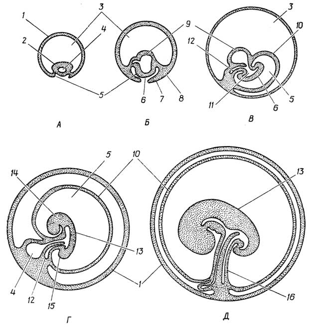
Fig. I.34. Human embryonic development:
A – late blastula at the time of embryonic implantation into the uterine wall, approximately on the 8th–10th day of development; B – gastrula at two weeks; C – embryo on the 20th day of development; D – embryo on the 25th day of development; E – embryo at one month.
1 – chorion membrane; 2 – inner cell mass; 3 – extraembryonic coelom; 4 – yolk sac; 5 – formation of the amniotic cavity; 6 – embryonic disc; 7 – ectoderm; 8 – mesoderm; 9 – endoderm; 10 – amnion membrane; 11 – archenteron cavity; 12 – allantois; 13 – developing embryo; 14, 15 – upper (cephalic) and lower parts of the future fetus; 16 – umbilical cord
of the ectoderm until the blastocoel is completely obliterated. The newly formed cavity—the gastrocoel (archenteron)—opens to the outside via the blastopore precisely at the site where invagination occurred. The cells of the inner layer of the gastrula form the endoderm.
Embryos of the most primitive animals, such as cnidarians, consist solely of the ectoderm and endoderm. In the embryos of all other animals, a third cell layer—the mesoderm—arises through various mechanisms, becoming localized between the ectoderm and endoderm.
The human embryo (Fig. I.34), referred to as a blastocyst following the blastula stage, differentiates into a hollow sphere, the outer envelope of which is termed the chorion, and a much smaller, dense cell mass attached to the inner surface of the chorion. Approximately on the tenth day after fertilization, the embryo attaches to the uterine wall, a process known as implantation. During the gastrula stage, the cells of the dense inner mass form two cavities: the amniotic cavity, surrounded by the ectoderm, and the yolk sac, lined by the endoderm. The cells of the germ disc lie between them. Further development proceeds such that the amniotic cavity gradually envelops the growing embryonic disc as well, which in turn encloses a portion of the yolk sac, shaping its remaining part into a long, slender tube. This tube subsequently forms the umbilical cord, connecting the fetus to the chorion, which at the same site externally forms the placenta together with the tissue of the uterine wall.
Table I.2
Embryonic origin of certain human organs
and tissues
Ectoderm |
Mesoderm |
Endoderm |
Upper skin layer (epidermis) and its derivatives, such as skin glands (sweat and sebaceous glands), receptor structures, hair |
Inner skin layer (dermis) |
Epithelium of the inner lining of the digestive and respiratory systems |
Nervous system, organs of vision and hearing |
Organs of the skeletal and muscular systems |
Liver and pancreas |
Epithelium of the oral and nasal cavities; receptor cells of the olfactory and gustatory organs |
Connective tissue and its derivatives, such as blood and blood vessels |
Epithelium of the inner lining of the urogenital system |
Adrenal medulla |
Kidneys and adrenal cortex, internal reproductive organs |
Thyroid and parathyroid glands |
Germ layers are three primary embryonic tissue types: the ectoderm, mesoderm, and endoderm. During fetal development, various organs are formed from these layers (Table I.2). Some organs originate from a single tissue type, whereas others involve multiple different tissues in their formation.
§ 4.
Basic Principles of
Animal Classification
As in the case of plants, the fundamental rule for grouping animals together is the commonality of their structure, development, and reproduction (Fig. I.35).
The characteristic used to divide all animals into two major groups—invertebrates and chordates—is the presence within the animal's body of a so-called notochord: a semi-rigid or fully flexible structure composed of cartilaginous or bony tissue that extends along the body and performs a skeletal support function.
Invertebrates are, in turn, subdivided into lower and higher invertebrates. Lower invertebrates possess discrete, still rudimentary organs that can be provisionally grouped into systems. Higher invertebrates feature a higher level of development across most systems, such as the digestive, circulatory, nervous, muscular, respiratory, excretory, and reproductive systems, as well as sensory and reproductive organ systems.
Chordates are subdivided into organisms with a primitive notochord and vertebrates. The former either have an insufficiently developed notochord or possess one only in the larval stage, with it disappearing in the adult organism; evolutionarily, they serve as a transitional link between invertebrates and vertebrates. The notochord of the latter is replaced by a cartilaginous (in lampreys, sharks, and rays) or bony (in most animals) vertebral column.
General Remarks
on Animal Classification
The most primitive animals are the cnidarians. They already possess all types of tissues, although their organs are quite simple: a digestive cavity with a single opening serving as both mouth and anus, tentacles, and uncomplicated reproductive organs (gonads). They still lack a mesoderm.
In free-living flatworms, a mesoderm is detected, and new organs appear: sensory organs, a muscular pharynx, and an excretory system composed of specialized cells. In parasitic flatworms, such as tapeworms, owing to the specific conditions of their habitat, only the reproductive organs are well developed, whereas the digestive cavity and mouth are absent since nutrients are simply absorbed through the body surface.
In nemerteans, roundworms, and other closely related taxa, organ systems emerge. A conventional digestive system with separate entrance and exit openings is present, nervous and muscular systems develop, and nemerteans feature a circulatory system containing hemoglobin. The refinement of the mesoderm leads to the formation of the coelom, i.e., a body cavity enclosed between two "tubes" —

Fig. I.35.
Principles of animal classification
|

the ectoderm (external surface) and the endoderm (digestive tract)—and filled with internal organs bathed in body fluid.
In higher invertebrates, organ systems undergo further refinement, giving rise to respiratory organs, endocrine "glands," and an exoskeleton. It is believed that evolution diverged from a common cnidarian ancestor into two main branches. One of these comprises flatworms, roundworms, annelids, mollusks, and arthropods, while the other led through echinoderms to chordates. A more distant ancestor of both animal branches, together with the ancestor of plants and fungi, originates from ancient protists, which in turn presumably descended from prokaryotes (Fig. I.36).
Section B
CELLS, TISSUES, AND ORGANS
IN ARTIFICIAL CONDITIONS
Chapter 6
KEY ASPECTS OF CULTIVATING
LIVING MATTER
§ 1.
Potentials of cell, tissue, and organ
culture in vitro
and associated challenges
Currently, maintaining the viability of living matter in vitro over extended periods is a challenge that has been successfully resolved for many specific cases.
The production of biomass from single-celled prokaryotic and eukaryotic organisms is widely used in both laboratory and industrial settings.
Technically somewhat more difficult, yet entirely feasible, is the in vitro cultivation of plant and animal tissues. An example of the former is the successful cultivation of ginseng root tissue culture or the regeneration of a complete carrot plant from phloem cells. An example of the latter is the cultivation of monkey kidney epithelial tissue to produce the polio vaccine. Tissue culture techniques currently make it possible to maintain their viability for very long periods (decades).
Even more challenging is maintaining the viability of organs isolated from the organism for a given period. Here, the primary task is to solve the problems of providing tissues and organs with nutrition.
As for plants, it is sufficient to place twig cuttings in water, since the uptake of nutrient solution (water and dissolved mineral substances) into plant organs is driven primarily by osmotic forces. Individual animal organs can be kept viable for many hours and even days under sterile and low-temperature conditions. The duration of viability for isolated animal organs depends not only on supplying their cells with nutrients, but also on how effectively metabolic waste products are removed from the organs.
§ 2.
Features of plant
and animal cell
cultivation
Two distinctive features emerge when growing specific cells or tissue samples in isolation from other tissues and organs.
The first feature—poor growth or its complete absence—is related to the fact that the development (i.e., proliferation and differentiation) of a particular cell type within an organism is normally regulated not only by substances the cells produce themselves, but also by the metabolic products of cells from other tissues and organs. For instance, the proliferation of myeloid stem cells (precursors of erythrocytes, platelets, macrophages, and mast cells) is driven by protein growth factors secreted by connective tissue fibroblasts.
Only embryonic and cancer cells multiply quite well under artificial conditions. The growth of the former is apparently due to the fact that under natural conditions, up to a certain stage, their development relies on internal reserves, and they do not require an external supply of nutrients and growth regulators. The latter cells are inherently characterized by uncontrolled growth that persists even in vitro. It is believed that the transformation of a normal cell into a cancer cell involves a reorganization of nuclear material, causing the cells to lose the ability to cease growth in a timely manner after a specified number of divisions. In other words, cancer cells remain in the proliferation stage indefinitely and do not transition to the final differentiation stage. They also lack contact inhibition, a mechanism that normally stops a cell from dividing once it comes into contact with neighboring cells of the same type.
Nerve cells are the most difficult to maintain in culture, especially when harvested from adult organisms. However, the growth of their neurites can be induced. This is achieved using nerve growth protein factors, which are synthesized by many tissues but are typically extracted from submandibular salivary glands, certain types of cancer cells, and snake venom.
The second feature is the rapid loss of differentiation capacity. When cultured, cells of any given tissue transform into one of three types: epithelial cells, fibroblasts, or amoebocytes. Epithelial cells are formed during the cultivation of epithelial tissues of both ectodermal and endodermal origin. Fibroblasts (mechanocytes) originate from muscle tissue cultures and various types of connective tissue. Amoebocytes, much like fibroblasts, are of mesodermal origin. They arise in cultures of macrophages and microphages.
Nerve cells and lymphocytes do not undergo transformation in culture.
§ 3.
Cloning —
a method for obtaining cultures
of genetically identical cells
A clone is a colony of cells derived by division from a single ancestral cell. Cloning is the technique of growing a cell clone; it is used when there is a need to obtain a culture of genetically identical cells.
§ 4.
Production of hybridomas —
a method for cultivating cell cultures
with predefined properties
A hybridoma is a cell produced by the fusion of two cells of completely different origins, such as a mouse cell and a human cell. Cell fusion in culture is achieved by treating cell membranes with chemical or physical methods (electrofusion). The hybridoma method is used when it is necessary to combine the properties of two cells of different origins within a single cell—for instance, the ability of lymphocytes to produce protective protein antibodies combined with the ability of cancer cells to grow robustly in culture.
Part II
STATIC BIOCHEMISTRY
Section A
PROTEINS —
THE PRIMARY MATERIAL OF THE CELL
Proteins account for up to half or more of the dry mass of an animal cell.
Chapter 7
ELEMENTAL COMPOSITION
AND PRIMARY STRUCTURE OF PROTEINS
The elemental composition of proteins includes C, O, H, N, and occasionally S. Proteins may also contain trace elements such as Fe, Cu, Zn, and others. However, the chemical elements themselves are not the "alphabet letters" that make up the vast diversity of protein "words" and molecules. Instead, nature chose simple compounds—α-amino acids—as the fundamental building blocks of proteins.
§ 1.
Building Blocks of Proteins: Amino Acids
All proteins are primarily composed of 20 α-amino acids (strictly speaking, one of them, proline, is an α-imino acid). Amino acids are generally divided into three groups based on their state at neutral pH (Fig. II.1). In addition to the standard amino acids listed in Fig. II.1, proteins can contain "unusual" amino acids, which are derivatives of standard amino acids (Fig. II.2), and prosthetic groups. These groups can vary greatly in composition and structure—from simple metal atoms or phosphate groups to rather complex molecules such as lipids, polysaccharides, and nucleic acids.

Fig. II.1 Protein amino acids at pH 7. Parentheses indicate the pK values (deprotonation constants) of the respective groups along with standard abbreviations.
§ 2.
Simple and Conjugated Proteins
Based on their composition, proteins are classified into simple and conjugated types. Simple proteins consist solely of amino acid residues, whereas conjugated proteins contain a non-protein prosthetic group in addition to amino acids.
Table II.1
Prosthetic groups of various
conjugated proteins
Type of Conjugated Protein |
Prosthetic Group |
Metalloproteins |
Metal atoms |
A conjugated protein that has lost its prosthetic group is referred to as an apoprotein. Conjugated proteins are further divided into classes (Table II.1) depending on the composition and structure of their prosthetic group.


Fig. II.2. Some "unusual" amino acids found in proteins
§ 3.
Properties of Amino Acids
As shown in Table II.2, at neutral pH values, amino acids exist as zwitterions—meaning their amino and imino groups carry a positive charge, while their carboxyl groups are negatively charged. This explains why amino acid molecules exhibit large dipole moments and their solutions possess high dielectric constants. The relatively high melting points of amino acids indicate that their crystals feature ionic lattices.
Aromatic amino acids absorb ultraviolet light, with tryptophan showing the strongest absorption (280 nm), followed by tyrosine (280 nm), and phenylalanine being the weakest (260 nm).
All amino acids, with the sole exception of glycine, possess a chiral center (an asymmetric carbon atom).
Table II.2
pKa values of various amino acid groups
Ionizable amino acid group |
pKa |
α-Carboxyl group |
2 |
Two amino acids—threonine and isoleucine—contain a second chiral center as well. To distinguish between stereoisomers, L- and D-glyceraldehyde have been universally adopted as configurational standards (Fig. II.3).

Fig. II.3. L-Glyceraldehyde (left) and D-glyceraldehyde (right)
Accordingly, amino acids also occur as L- and D-stereoisomers (Fig. II.4). The corresponding projection formulas are

These formulas represent a two-dimensional projection of what an observer sees when viewing the stereoisomers from the direction indicated by the arrows in Fig. II.4. The DL system of stereoisomer nomenclature remains standard for amino acids and sugars. The more general RS system coincides with the DL system in most cases
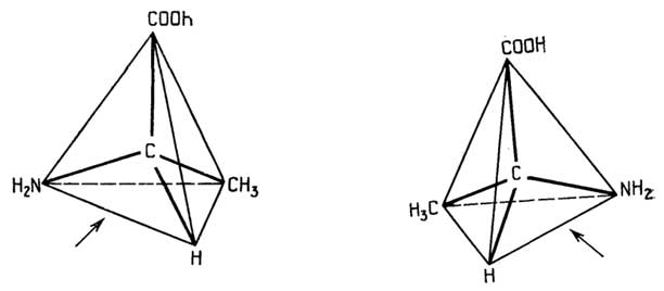
Fig. II.4. L-Alanine (left) and D-alanine (right)
(especially for amino acids and sugars). All natural proteins and peptides synthesized on ribosomes typically consist of L-amino acids.
§ 4.
The Basis of Protein Structure:
The Polypeptide Chain
Amino acids form polymer chains via peptide bonds:
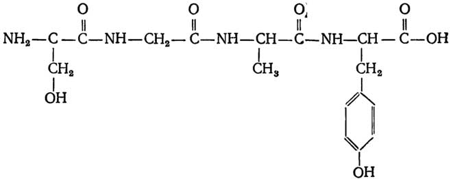
When writing amino acid sequences in shorthand, the N-terminus is written on the left and the C-terminus on the right.
The peptide bond is characterized by a high degree of stabilization due to resonance:

This results in the following: 1) the imino group is incapable of accepting or releasing a proton within the pH range from 0 to 14; 2) the bond, rather than being a pure single bond, is "rigid," meaning free rotation around it is impossible.
Sturally, the atoms of the peptide bond lie in nearly the same plane, with the nitrogen-bonded atoms forming a pyramid with an obtuse apex at the nitrogen atom (Fig. II.5). The atoms

Fig. II.5. Spatial arrangement of atoms in a peptide bond
of oxygen and hydrogen are most commonly in the trans-position, although the somewhat less stable cis-peptide bond (involving proline) also occurs.
Through peptide bonds, the body forms both simple peptides (di-, tri-, and oligopeptides) and polypeptides—that is, proteins with amino acid residue counts ranging from fifty to several thousand. A protein molecule may consist of one or more polypeptide chains. An individual polypeptide chain contains anywhere from several dozen to several hundred amino acid residues. Polypeptide chains are unbranched, but they may be covalently cross-linked internally or with one another through the formation of disulfide bonds between spatially proximate Cys SH-groups:
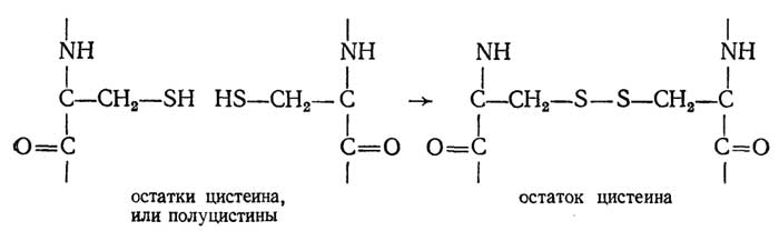
The sequence of amino acid residues in a polypeptide chain, taking into account "cystine bridges," constitutes the primary structure of proteins. It is worth noting that this structure is maintained by covalent bonds.
Chapter 8
SPATIAL STRUCTURE OF PROTEINS
Based on their spatial structure, proteins are divided into globular and fibrous.
Globular proteins consist of one or more polypeptide chains tightly folded, via non-covalent and frequently covalent bonds, into a compact particle known as a globule. They are generally readily soluble in water. Enzymes, antibodies, many hormones, and transport proteins—such as myoglobin and hemoglobin (oxygen carriers) or serum albumin (fatty acid carrier)—are all globular proteins.
Fibrous proteins consist of elongated or helical polypeptide chains arranged parallel to one another and held together primarily by numerous non-covalent and occasionally covalent bonds. These polypeptide chains are assembled into fibers (fibrils). Such proteins are insoluble in water. Examples include hair, nails, feathers (keratins), tendons (collagen), ligaments (elastin), silk, and spider silk (fibroin).
The differences between the structure of globular and fibrous proteins, as well as their properties (such as solubility), are evident.
§ 1.
Primary Structure Determines
the Spatial Conformation of a Protein
The principle stated in the heading was experimentally confirmed by C. Anfinsen. He disrupted the native three-dimensional structure of the enzyme ribonuclease (denatured it) by adding urea and β-mercaptoethanol to the protein solution. Then, by removing the denaturing agents via dialysis, he restored the active protein (renatured it). Thus, the polypeptide chain folds into its "native" structure spontaneously, driven by factors inherent in the structure of the polypeptide chain itself.
There are several reasons why a given polypeptide chain adopts a single three-dimensional structure—its native conformation. Within the polypeptide chain (Fig. II.6), we observe

Fig. II.6. Alternation of the "rigid" peptide bond and single bonds in a polypeptide chain
repeated alternation of bonds: peptide and two single bonds. Theoretically, free rotation of the planes containing "rigid" peptide bonds around the single bonds is permissible. Their deviations from the planes of the figure are determined by the angles φ and ψ. However, in reality, a whole range of restrictions is imposed on the possible values of φ and ψ.
Constraints associated with interactions within the chain itself
Steric hindrances. Simultaneous free rotation around different single bonds is impossible, as can be verified using molecular models. Polyglycine possesses the highest number of degrees of freedom. An increase in the size of the side chains R reduces the possibility of free rotation around single bonds.
Dipole interactions. The plane of a peptide bond constitutes a dipole, which naturally tends to orient itself relative to other dipoles in a specific manner, especially if this arrangement is stabilized by the formation of bonds, such as hydrogen bonds.
Interaction of R side chains. Positive and negative charges or the hydrophobicity of adjacent side chains hinder or promote a specific spatial arrangement of the peptide planes.
Constraints associated with chain interactions
with the environment
(with the solvent or other neighboring chains):
formation of hydrogen bonds;
hydrophobic interactions;
ionic interactions;
formation of cystine bridges.
All these constraints result in the polypeptide chain adopting only a single native conformation.
Levels of spatial structure of proteins
The spatial structure of proteins can be disordered, i.e., a statistical coil like that of a denatured protein, or ordered, like that of a native protein. In the latter case, it is customary to consider:
secondary structure — the spatial organization of amino acid residues adjacent in the chain;
tertiary structure — the spatial organization of amino acid residues distant in the chain;
quaternary structure — the spatial arrangement in a single molecule of individual polypeptide chains, each folded into its own globule. Such globules are called protomers or subunits. The connection between them in a protein molecule is provided by non-covalent forces. Proteins whose molecules consist of several subunits (protomers) are called oligomeric.
Tertiary and quaternary structures are typically attributed to globular proteins. In fibrous proteins, the boundary between the levels of spatial organization is somewhat conventional.
§ 2.
Secondary structure of proteins
The first successful attempt to construct a regular secondary structure for a polypeptide chain was made by Pauling and Corey in 1951. Based on a comparison of possible spatial arrangements of polypeptide molecular models, they concluded that they exist in two forms: the α-helix and the β-pleated sheet, which was experimentally confirmed a few years later. Subsequently, short segments of other helices were discovered in globular proteins, which were designated as the 310-, π-, and αII-helices, respectively.
Types of secondary structures
The right-handed α-helix is the most common helical structure of polypeptide chains formed by L-amino acids in proteins (Fig. II.7). The spatial arrangement of amino acid residues within it determines the most stable structure. Stabilization is achieved due to the fact that the turns of the helix are "stitched" by hydrogen bonds, as well as by dipole interactions. The number of hydrogen bonds is maximal. Their orientation is almost parallel to the longitudinal axis of the α-helix. There are 3.6 amino acid residues per turn, and all peptide bond dipoles are directed downwards. The number of atoms in the ring formed by the hydrogen bond is 13. The hydrogen bond arises between the CO group of the n-th residue and the NH group of the (n + 4)-th residue (Fig. II.8), i.e., interaction of closely spaced and unidirectionally oriented peptide bond dipoles occurs every two dipoles.
The 310-helix (Fig. II.9) is a right-handed helix, more tightly wound than the α-helix. The accepted designation 310 is related to the fact that the helix is characterized by three residues per turn and 10 atoms in the ring formed by the hydrogen bond. The latter arises between the CO group of the n-th residue and the NH group of the (n + 3)-th residue (Fig. II.10), i.e., in this helix, the dipoles are closer together and interact with each other across one.
The π-helix (Fig. II.11) is characterized by 4.4 amino acid residues per turn and a larger diameter than the α-helix. When the hydrogen bond is formed, a 16-atom ring is produced (Fig. II.12). Thus, the π-helix can be designated
Fig. II.7. Scheme of a right-handed α-helix. Dashed lines indicate hydrogen bonds, arrows indicate peptide bond dipoles. Dots indicate amino acid residues located on the invisible side of the cylinder |
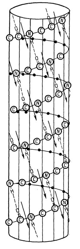

Fig. II.8. Formation of hydrogen bonds in the α-helix. For clarity, the peptide bond dipoles (top) and amino acid residues (bottom) are numbered;
|
Fig. II.9.Diagram of the right-handed 310-helix. Hydrogen bonds are indicated by dashed lines, and peptide bond dipoles by arrows. Dots denote amino acid residues located on the back side of the cylinder |

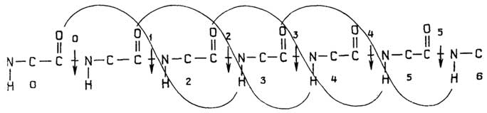
Fig. II.10.Formation of hydrogen bonds in the 310-helix
Fig. II.11.Diagram of the right-handed π-helix. Hydrogen bonds are indicated by dashed lines, and dipoles by arrows. Dots denote amino acid residues located on the back side of the cylinder |
|

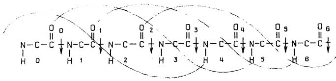
Fig. II.12.Formation of hydrogen bonds in the π-helix
|
Fig. II.13.Diagram of the right-handed αII-helix. Dashed lines indicate hydrogen bonds, and arrows indicate peptide bond dipoles. Dots represent amino acid residues on the back side of the cylinder |


Fig. II.14.Formation of hydrogen bonds in the αII-helix
as a 4.416-helix1. The hydrogen bond is formed between the CO group of the n-th residue and the NH group of the (n + 5)-th residue, while the dipoles approach and attract each other across three residues, i.e., the 1st to the 5th, the 2nd to the 6th, etc.
|

The αII-helix (Fig. II.13) has 4 residues per turn in a cycle with a 14-atom hydrogen bond (Fig. II.14). The hydrogen bond is formed between the NH group of the n-th residue and the CO group of the (n + 3)-th residue, and the dipoles interact with each other across three residues.
Comparing the αII-helix with those already listed, characteristic differences can be observed:
all dipoles are directed upward;
hydrogen bonds are formed between the beginning (NH group) of the n-th residue and the end (CO group) of the (n + 3)-th residue2.
The collagen helix (Fig. II.15) is found exclusively in collagens—the most abundant proteins in humans and higher animals. They serve as the structural basis for tendons, cartilage, skin, bones, etc. The polypeptide chain of collagen forms an irregular, left-handed, and rather extended helix with three amino acid residues per turn. There are no intrachain hydrogen bonds within the helix. Stabilization is achieved through interchain covalent bonds, as well as hydrogen bonds and hydrophobic interactions (see p. 121). The polypeptide chain contains approximately 1000 residues, with a molecular weight of 100,000.
Fig. II.15.Diagram of the collagen helix. Small circles denote amino acid residues forming a left-handed helix, which in turn twists into a right-handed superhelix
Features of collagen composition:
one-third of the composition is glycine;
one-fifth is proline and its hydroxy derivatives;
other amino acids are present in small quantities and in an incomplete set;
presence of the rare amino acid 5-hydroxylysine.
The exact structure of the elastin helix—the main protein of bone ligaments—remains unknown. It contains about 800 residues; molecular weight is 72,000.
Features of the elastin chain composition:
one-third of the composition is glycine;
high proline content (one-eighth), but low hydroxyproline content;
half of the composition consists of hydrophobic aliphatic amino acids Ala, Val, Leu;
other amino acids are contained in small quantities and in an incomplete set;
repeated sequences are frequently encountered, such as Pro-Gly-Val-Gly-Val, Lys-Ala-Ala-Lys.
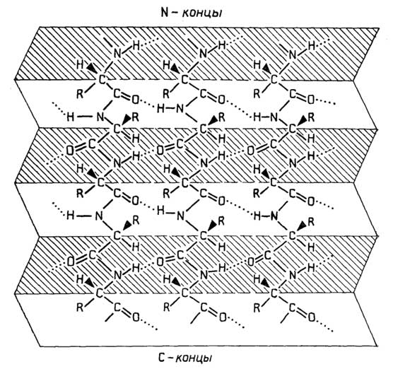
Fig. II.16. Parallel β-pleated sheet. The N-termini of the chains are at the top, and the C-termini are at the bottom.
The β-pleated sheet is formed by fairly extended polypeptide chains. There are two types:
parallel – when the polypeptide chains run in the same direction (Fig. II.16);
antiparallel – when the polypeptide chains run in opposite directions (Fig. II.17).
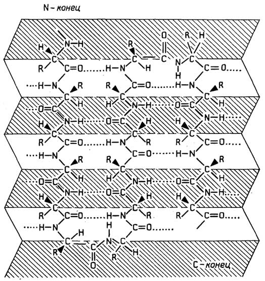
Fig. II.17. Antiparallel β-pleated sheet. A single chain.
Its N-terminus is at the top left, and the C-terminus is at the bottom right. The chain
turns to form two β-turns.
The stabilization of β-pleated sheets is achieved through the formation of interchain hydrogen bonds involving all peptide bonds.
Features of β-pleated sheets:
the chains are extended, but not fully, resulting in a "corrugated" structure rather than a flat plane;
hydrogen bonds lie within the planes of the pleats;
R-groups are arranged in rows, alternately above and below the plane of the drawing;
an antiparallel β-pleated sheet can be formed by a single polypeptide chain that folds back on itself to form β-turns.
The secondary structure is determined by the primary structure. This is evidenced by X-ray diffraction data of synthetic polypeptides as well as proteins with known amino acid sequences. Based on these data, the 20 amino acids can be classified quite strictly according to their propensity to form helices or β-structures
Table II.3
Propensity of protein L-amino acids to form helices
and β-structures
Helices |
β-Structures |
|
Strong formers |
Glu, Ala, Leu |
Val, He, Met |
Formers |
His, Gin, Val, Phe, Trp, Met |
Thr, Tyr, Cus, Gin, Leu, Trp, Phe |
Weak formers |
Lys, Ile |
Ala |
Indifferent |
Asp, Arg, Ser, Thr, Cys |
Asp, Arg, Gly |
Strong breakers |
Asn, Tyr |
His, Lys, Ser, Asn, Pro |
Breakers |
Gly, Pro |
Glu |
into several groups (Table II.3). Using these data for the theoretical prediction of secondary structure has yielded good results in a number of cases.
§ 3.
Tertiary Structure
of Globular Proteins
Analysis of spatial structure data for proteins with known architectures leads to the conclusion that globular proteins can be divided into five groups based on their tertiary structure.
Types of Globular Architecture
α-Proteins – proteins with a high content of helical structures, such as myoglobin, an oxygen-binding muscle protein. It releases stored oxygen as needed to support metabolism. It is typically isolated from the muscles of diving animals such as whales and seals. Myoglobin was the first globular protein whose tertiary structure was elucidated by X-ray diffraction analysis (Fig. II.18). The myoglobin molecule consists of a single polypeptide chain of 153 residues, with a molecular weight of 16,700.

Fig. II.18. Myoglobin:
A – schematic representation of the tertiary structure of myoglobin; the hatched area represents the heme group, with the dark spot indicating the Fe atom; B – structure of heme (Fe-protoporphyrin IX); C – oxymyoglobin.
Structural features of myoglobin:
about 75% of its polypeptide chain is in the α-helical conformation, comprising several helical segments;
between the helical segments lie amino acid residues that disrupt the α-helix, such as Pro, "charged" residues Asp, Lys, Arg, as well as Ser, Thr, Tyr capable of forming hydrogen bonds with carboxyl groups;
hydrophobic residues are hidden inside the molecule, while polar and "charged" residues are located on the outside;
only four water molecules can fit inside the molecule;
the heme prosthetic group is non-covalently bound to the apoprotein.
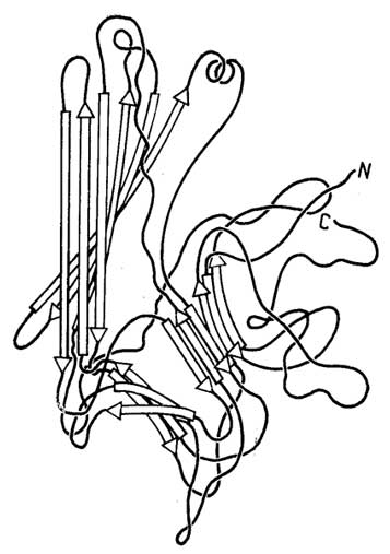
Fig. II.19. Diagram of the tertiary structure of a concanavalin A subunit globule, consisting of antiparallel β-sheets. The propeller-like twisting of the β-sheets is clearly visible.

Fig. II.20. Diagram of the tertiary structure of a bovine erythrocyte superoxide dismutase subunit globule.
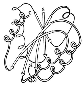
Fig. II.21. Diagram of the tertiary structure of a flavodoxin globule. A parallel β-sheet is flanked by two α-helices above and three below. A non-covalently bound flavin mononucleotide (FMN) is located between two β-segments (see Fig. II.19).
β-Proteins: their globules consist of two or more folded β-sheets. Such proteins are relatively rare. An example is the plant-derived oligomeric protein concanavalin A. Its molecule consists of four subunits, each representing a polypeptide of 237 residues with a molecular weight of 27,500 (Fig. II.19). Concanavalin A belongs to a group of proteins collectively known as lectins. These proteins are characterized by the ability to specifically interact with
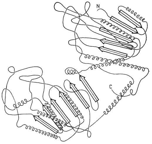
Fig. II.22. Diagram of the tertiary structure of phosphoglycerate kinase.
The α-helix regions are located above and below the β-sheets.
the cell surface, causing, for example, cell agglutination. Concanavalin A itself agglutinates cancer cells and is also capable of inducing lymphocyte proliferation.
Another example of a β-protein is bovine erythrocyte superoxide dismutase (SOD). Its molecule consists of two subunits, each being a polypeptide of 151 residues with a single cystine bridge. The structure of the SOD subunit is also based on an antiparallel β-sheet (Fig. II.20), although in this case it is rolled into a barrel shape.
α/β-Proteins: the polypeptide chain consists of alternating α-helices and extended β-segments grouped into a single β-sheet, as seen, for example, in bacterial flavodoxin or
phosphoglycerate kinase. The flavodoxin molecule contains 138 residues. Five segments of the chain form a parallel propeller-twisted β-sheet with α-helices on either side (Fig. II.21). The phosphoglycerate kinase molecule consists of 355 residues and comprises two β-sheets with numerous helices on both sides (Fig. II.22).

Fig. II.23. Diagram of the tertiary structure of the thermolysin molecule. The pleated β-sheet consists of 10 parallel and antiparallel strands. Seven helical regions are visible.
(α + β)-Proteins: the polypeptide chain can be divided into regions consisting entirely of helices and regions having a pleated β-sheet conformation. Such a structure exists in the thermolysin molecule (Fig. II.23), whose polypeptide chain contains 316 residues.
(β, β)-Proteins are virtually devoid of helical and pleated structures. An example is ferredoxin from Peptococcus aerogenes (Fig. II.24). At first glance, the polypeptide chain of such proteins appears to fold into a globule as a random coil, but this is not the case. The same forces that operate in other proteins govern the folding of the globule.
Domains
Molecular globules (or subunit globules) sometimes consist of clearly distinguishable lobes, as seen in the examples of phosphoglycerate kinase (see Fig. II.22) and thermolysin (see Fig. II.23). Such covalently linked lobes are commonly referred to as domains. Domains are a characteristic feature of the tertiary structure of certain globular proteins.
A comparison of the tertiary structures of a number of proteins has led to interesting conclusions. First, proteins with similar functions but different primary, secondary, tertiary, and quaternary
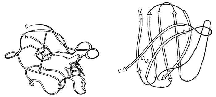 G">
G">
Fig. II.24. Diagram of the tertiary structure of the ferredoxin protein from Peptococcus aerogenes. Dark circles represent iron atoms, and light circles represent sulfur atoms.
Fig. II.25. Diagram of the tertiary structure of the variable domain of the immunoglobulin G heavy chain.
structures often contain similar domains. For example, many dehydrogenases, which require nicotinamide adenine dinucleotide (NAD) for their function (see Fig. II.18), share a common NAD-binding domain. Second, proteins with different functions and structures sometimes also contain structurally similar domains. For example, one of the domains (the variable domain) of the immunoglobulin G heavy chain (Fig. II.25) bears a striking spatial resemblance to the subunit globule of bovine erythrocyte superoxide dismutase (see Fig. II.20).
Nature of the forces stabilizing the tertiary
structure of proteins
The stabilization is primarily driven by hydrophobic interactions between the side chains of amino acid residues, which group together inside the globule to avoid thermodynamically unfavorable contact with water molecules. Hydrogen bonds play an equally important role. They form both between amino acid side chains and between the NH and CO groups of peptide bonds. The electrostatic attraction of opposite charges on side chains—such as COO- and NH3+—is also significant. The strongest bonds, although present in only some proteins, are covalent cystine bridges.
Factors causing
protein denaturation
The weakening of one or more of the aforementioned interactions, as well as the cleavage of S–S bonds, can lead to the unfolding of the polypeptide chain—that is, the disruption of the native tertiary structure of the protein, known as denaturation. Denaturation is caused by:
extreme pH values that disrupt electrostatic interactions and break hydrogen bonds;
temperature, which primarily breaks weak non-covalent bonds;
high salt concentrations, which also disrupt electrostatic interactions and break hydrogen bonds;
urea, guanidine, and organic solvents, which break hydrophobic interactions as well as hydrogen bonds;
sulfhydryl compounds such as β-mercaptoethanol, other reducing agents like NaBH4, and oxidizing agents such as performic acid, which cleave cystine bridges (the latter are not always accessible if buried inside the globule).
Denaturation can be irreversible or reversible. Irreversibility can often be explained by kinetic factors.
Formation of the native structure
The formation of the native structure is a cooperative process. The formation of the α-helix itself is thermodynamically favorable if a sufficient number of turns "stitched" together by hydrogen bonds are formed. When a single turn is formed, a rather high entropic barrier must be overcome, as a single hydrogen "stitch" entails the loss of rotational freedom for six bonds; however, each subsequent adjacent "stitch" restricts the rotational freedom of only two more bonds (Fig. II.26). Thus, in the equation ∆G = ∆H - T∆S for α-helix formation, the negative value of ∆S is more than compensated for by the equally negative value of ∆H for hydrogen bond formation, and the more hydrogen bonds formed, the more thermodynamically favorable the α-helix becomes. A significant contribution to helix stabilization (more precisely, to the negative value of ∆H) is also made by dipole-dipole interactions (see Figs. II.7, II.9, II.11, II.13). Calculations show that these interactions are most fully manifested in a helix of ten residues.

Fig. II.26. The formation of a single hydrogen bond in an α-helix leads to the loss of rotational freedom of six bonds (indicated by arrows) located inside the turn, whereas the formation of the second hydrogen bond restricts only two bonds
The coil-helix transition occurs as follows: first, a single turn with a single hydrogen bond acting as a nucleus appears in a segment of the polypeptide chain known as the nucleation center. This is highly likely to occur where four adjacent residues belong to the category of "helix-promoting" residues. Then, starting from the initial turn, helix propagation accelerates in both directions along the chain until it encounters residues that "disrupt helix formation."
The formation of β-sheets is analogous to helix formation, with the fundamental difference that hydrogen bonds form between separate polypeptide chains. The role of the "nucleus" is played by chain segments consisting of three residues that actively promote β-sheet formation.
The formation of the native protein conformation occurs through the association of α-helices and β-structures. Calculations show that forming the native conformation by simply testing all possible variants and selecting the most energetically favorable one is completely unrealistic, as it would take many billions of years. A compelling hypothesis suggests that it happens as follows: small segments (10–15 residues) of helices and β-structures, even if short-lived, approach each other via diffusion and are stabilized by forming aggregates called "folding units." Subsequent formation of the native structure proceeds with acceleration.
In proteins poor in helices and β-structures, other chain segments and factors—such as complexation with metal ions—likely play an equally important stabilizing role. Such a process, where each subsequent step is facilitated and accelerated, is called cooperative. Only through cooperativity can protein renaturation occur rapidly enough under appropriate conditions.
§ 4.
Quaternary Structure
of Globular Proteins
In many proteins, the molecule consists of more than one globule.
Hemoglobin is an example of an allosteric protein
Structure of hemoglobin. The hemoglobin molecule in vertebrates consists of four subunit globules (protomers). For example, hemoglobin A (the main human hemoglobin) consists of two identical α-subunits (141 residues each) and two identical β-subunits (146 residues each). Its subunit formula is α2β23, with a molecular mass of 64,500. The four protomers are arranged in a tetrahedron (Fig. II.27). Permanent bonding between them is maintained by hydrophobic interactions. Each α-protomer has extensive contact with both β-protomers. Contact between identical protomers is weaker. The hemoglobin molecule easily dissociates into two αβ-halves. Further dissociation occurs under harsher conditions. From this, it can be concluded that the interactions between α and β' and between α' and β are substantially weaker than the interactions between α and β, as well as between α' and β'. Each protomer contains a heme located in a hydrophobic "pocket," which protects it from oxidation to the ferric form.
The heme groups are located on the outer surface of the protomers. The distances between them are quite large—2.5 nm. The hemoglobin molecule exists in two forms: the "tense" (T) and the "relaxed" (R) state. The former is characteristic of
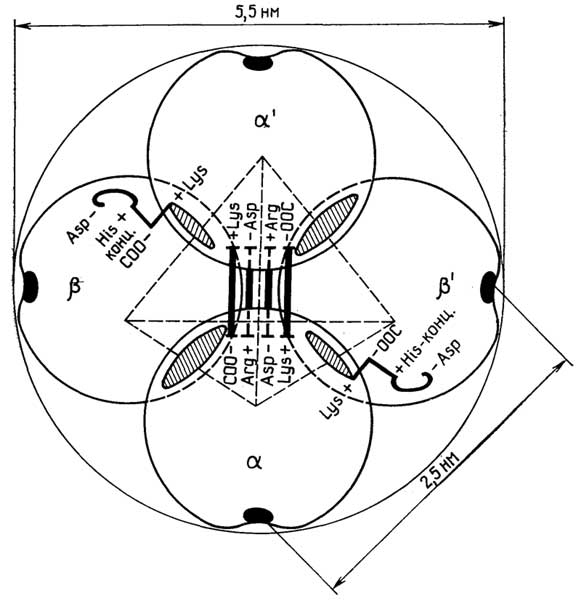
Fig. II.27. Diagram of the quaternary structure of the hemoglobin molecule. Hatched areas indicate contact sites of α- and β-protomers, black areas indicate heme locations, and bold lines show "salt bridges" present in deoxyhemoglobin. There is a small cavity in the center of the tetrahedron
deoxyhemoglobin, while the second is characteristic of oxyhemoglobin. The T-form is more "rigid"; unlike the R-form, it possesses eight salt bridges:
four salt bridges between the α-protomers;
two salt bridges between different protomers, specifically between α and β' and between α' and β;
one salt bridge within each β-protomer.
The formation of four salt bridges involves the C-terminal carboxyl groups and the side-chain amino groups of four lysines located inside the α-chains. The formation of the other four salt bridges involves the side-chain carboxyl groups of four aspartic acid residues on one side, and the side-chain groups of two arginines and two histidines on the other.
Upon oxygenation, the salt bridges are broken, and the T-form transitions into the more compact R-form. In this process, one αβ-half rotates by 15° and shifts by 0.08 nm relative to the other. This entails virtually no structural changes in the globules at the contact sites of the α and β, as well as α' and β' protomers. However, at the contact sites of the α and β', as well as α' and β protomers, the globule structure changes to such an extent that some atoms in these regions shift by distances of up to 0.6 nm.
Hemoglobin protomers are similar in structure and function to the myoglobin globule. A comparison of the α- and β-protomers of hemoglobin, as well as myoglobin in a single vertebrate species, shows that:
the primary structures of the α- and β-protomers differ considerably: 30–40% of the positions in the polypeptide chains are occupied by the same residues, which are referred to in such cases as invariant or conserved residues;
in terms of primary structure, the α- and β-chains of hemoglobin differ even more from the single chain of myoglobin, sharing only 20% invariant residues;
the tertiary structures of the α- and β-protomers of hemoglobin are practically identical;
the tertiary structure of myoglobin differs very little from the tertiary structures of the hemoglobin α- and β-protomers.
|
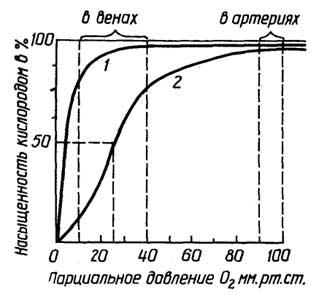
A comparison of hemoglobins and myoglobin across various animal species and humans demonstrates that while their primary structures differ significantly (with only 9 invariant residues), their tertiary structures exhibit a remarkable similarity that can only be explained by the commonality of their functions.
The functional features of hemoglobin are determined by its
Fig. II.28. Oxygen saturation curves for myoglobin (1) and hemoglobin (2). Partial pressure of oxygen in arteries is 90–100 mmHg, and in veins, 10–40 mmHg.
quaternary structure. The functional role of hemoglobin is the same as that of myoglobin—to reversibly bind molecular oxygen. Although each hemoglobin protomer binds oxygen in exactly the same way as myoglobin, the functioning of the hemoglobin molecule reveals significant differences: first, oxygen is bound cooperatively by the hemoglobin molecule, meaning that the binding of one O2 molecule facilitates the binding of the next (Fig. II.28), which results in more efficient oxygen transport. This type of cooperativity is termed positive cooperativity, in contrast to negative cooperativity, where the binding of one molecule impedes the binding of subsequent ones. Second, the hemoglobin molecule is capable of receiving "information" from its environment and, as a consequence, altering its affinity for oxygen; for example, protons and CO2 molecules (whose concentration is elevated in working muscles), by binding to hemoglobin at sites quite distant from the hemes, weaken its bond with oxygen (the Bohr effect). The organic phosphate 2,3-diphosphoglycerate (DPG) acts in a similar manner:
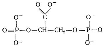
An increase in its concentration within erythrocytes occurs in various instances of oxygen deprivation, such as when a person ascends to high altitudes.
These features of hemoglobin stem from its quaternary structure, as they are absent in protomers, αβ-subunits, and hemoglobin β4 (hemoglobin H). Proteins whose affinity for a functionally essential molecule (substrate) is regulated by the binding of various effectors at other sites of the protein molecule are called allosteric.
Mechanism of allosteric
regulators' action
using hemoglobin as an example
This is explained by conformational changes within the protein molecule. When an O2 molecule binds to the Fe atom of the heme group, it pulls the iron into the plane of the heme (Fig. II.29). The proximal histidine follows the Fe atom, which in turn induces conformational changes in the protomer contact areas via other atoms. Thus, information about O2 binding by the heme of one protomer is transmitted to another protomer through their contact. This conformational rearrangement leads to the disruption of salt bridges, stabilization of the R-form, and, consequently, an increase in hemoglobin's affinity for oxygen.
Protons reversibly bind to the side chains of histidines at the C-termini of β-protomers and to the α-amino groups at the N-termini of α-protomers, as well as, potentially, to the side chains of two other histidines within the α-protomers.
CO2 molecules reversibly bind to the α-amino groups of the N-termini of α- and β-protomers.
DPG reversibly binds via electrostatic interactions to the positively charged N-termini of β-protomers, thereby holding the latter together.

Fig. II.29. Displacement of the proximal histidine upon oxygenation of deoxyhemoglobin
Protons, CO2, or DPG bind exclusively to the T-conformation (deoxyhemoglobin), providing additional stabilization. Upon the oxygenation of even a single protomer, conformational changes are transmitted to the other protomers, and the affinity for H+, CO2, and DPG decreases both for the first oxygenated protomer and for the hemoglobin molecule as a whole.
Other proteins
with complex quaternary structure
Proteins whose quaternary structure consists of several or dozens of subunits are generally referred to as oligomeric. Protein structures comprising hundreds or even thousands of subunits will be termed polymeric protein

Fig. II.30. Shapes and sizes of certain viruses:
A - spherical coat of the smallpox virus; B - common cold adenovirus, whose coat consists of 252 subunits; C - tadpole-shaped bacteriophage T4; D - tobacco mosaic virus (TMV) rod, whose coat consists of 2,200 subunits
complexes (PCs). The division of proteins into oligomers and PCs is quite arbitrary.
The simplest case of oligomers is when a protein molecule consists of two identical protomers. However, oligomers with up to several dozen subunits are also found, with the molecule simultaneously containing both identical and
different protomers, for example, RNA polymerase (mol. wt. 400,000) α2ββ'σ or the massive pyruvate dehydrogenase complex (mol. wt. 4,600,000) t24(p2)12(f2)12. Oligomers may have different symmetries, but they are always compact.
Polymeric protein structures can be broadly divided into two types based on the ratio of their length to diameter (or width).
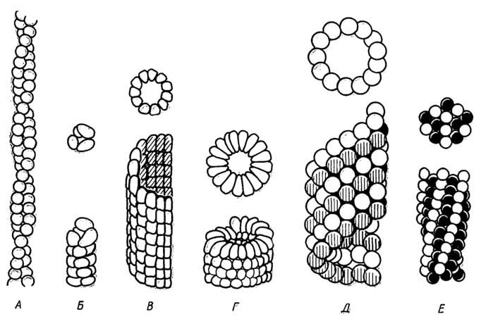
Fig. II.31. Subunit structure of certain complex proteins viewed from the end (top) and from the side (bottom):
A - muscleF-actin;
B - prokaryotic pilus;
C - prokaryotic flagellum;
D - tobacco mosaic virus coat protein;
E - microtubules;
F - deoxyhemoglobin S
Polymeric protein structures whose length is comparable to their diameter. Examples include the protein coats of spherical viruses, such as the smallpox virus; or those with a nearly polyhedral icosahedral shape, like the virus causing acute respiratory infections; or tadpole-shaped structures consisting of a polyhedral head with a relatively short cylindrical tail, as seen in bacteriophage T4 (Fig. II.30).
Polymeric protein structures whose length is significantly greater than their diameter, such as muscle F-actin, which accounts for about 25% of total muscle proteins. It forms a helix (Fig. II.31, A) 1000 nm long and 7 nm in diameter, twisted from two strands, each of which is a chain of non-covalently linked globular G-actin subunits (mol. wt. 46,000, diameter 2.4 nm, single polypeptide chain of 376 amino acid residues). This group of protein structures also includes: bacterial pili and flagella (Fig. II.31, B, C); the protein coat of the tobacco mosaic virus, which has the shape of long rods (Fig. II.31, D); microtubules (Fig. II.31, E), a constituent of the cytoskeleton as well as eukaryotic cilia and flagella; and deoxyhemoglobin S fibers (Fig. II.31, F), which are responsible for the hereditary disease sickle cell anemia.
The general principle underlying the assembly of such complexes is spiralization, which can be a single helix (bacterial pili, tobacco mosaic virus); a double helix (F-actin); or multistranded helices (prokaryotic flagella, microtubules, deoxyhemoglobin S fibers).
The Special Role of Quaternary Structure
Proteins with a quaternary structure evolved because:
they are better adapted to perform their functions due to their sensitivity to the regulatory action of the environment;
their biosynthesis saves genetic material and reduces the probability of errors.
§ 5.
General Principles of
the Spatial Structure
of Globular Proteins
During the folding of polypeptide chains into globules, as well as the formation of oligomeric or polymeric structures by the latter, several general patterns can be observed.
The formation of tertiary or quaternary structures occurs such that hydrophobic residues cluster inside the molecule, while polar and charged residues remain on the outside. This explains the solubility of globular proteins in water or aqueous salt solutions, as well as the stability of quaternary structures in these solutions.
Tertiary and quaternary structures are very compact. Only in certain cases can individual ions or water molecules penetrate their interior.
A native protein typically has one or, in some cases, several conformations. Such a native conformation is fairly stable under physiological conditions, but it is not entirely rigid. Its elements (atoms, atomic groups, domains, subunits) retain a small degree of freedom of spatial movement. During functioning, the native structure of many proteins undergoes a breathing-like motion.
§ 6.
Spatial Structure
of Fibrous Proteins
The fundamental difference between fibrous and globular proteins is that their polypeptide chains do not fold into globules; instead, remaining extended as linear chains or helices, they cluster into elongated fibers (fibrils). As in globular proteins, the basis of the spatial structure of fibrils is secondary structures: the α-helix, β-pleated sheet, and other types of helices.
α-Keratins
α-Keratins form the basis of hair, wool, feathers, nails, claws, horns, hooves, and turtle shells. The secondary structure is a right-handed α-helix. The structure of hair α-keratin is shown in Fig. II.32. Synthesized α-keratin forms fibrils that fill the entire cell. It is believed that protein molecules with a higher cystine content are located in the space between the fibrils. Subsequently, the cells die, and their walls transform into the hair sheath, or cuticle. Similar processes occur during the formation of α-keratins in other proteins.
Characteristics of α-Keratin
The polypeptide chains (in the form of α-helices) are parallel and oriented in the same direction.
The polypeptide chains of adjacent fibrils are cross-linked by covalent disulfide bonds between cystine residues. As a result, the fibers are firmly bound into a single entity. The higher the cystine content in α-keratin, the stronger and more rigid its structure (turtle shell contains 18% cystine).
The polypeptide chains contain a relatively large number of amino acids with hydrophobic side chains located on the outer surface of the α-helices, supercoils, and the fibrils themselves.
Flexible α-keratins with a low proportion of cystine (hair, wool yarn) can be stretched to double their length when treated with steam at high temperatures. This treatment breaks the intrachain hydrogen bonds; the α-helices straighten out, and a β-conformation forms, similar to that possessed by β-keratins (see below). The secondary structure of the β-conformation of α-keratins is a parallel β-pleated sheet. After the stretching load is removed and the material is cooled, the hair or wool α-keratin returns from the β-conformation to its original α-helical conformation. This is because α-keratin contains a relatively high number of amino acid residues with rather bulky side groups. The interaction of these groups destabilizes the parallel β-pleated sheet, making the β-conformation of α-keratin unstable.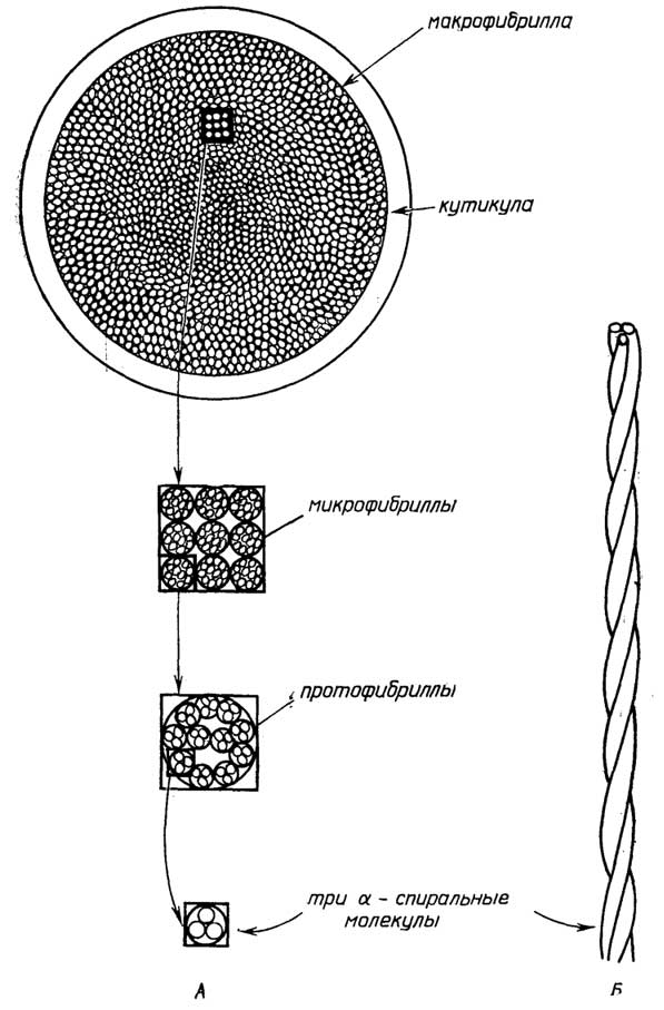
Fig. II.32. Structure of hair α-keratin. Three α-helical polypeptide chains are twisted into a supercoil, the protofibril. 11 such parallel protofibrils form a microfibril, which in turn assemble into macrofibrils.
β-Keratins
β-Keratins are proteins that form the fibers of silk and spider webs (fibroin). Their secondary structure is an antiparallel β-pleated sheet.
Characteristics of β-Keratins
Interchain covalent disulfide bonds are absent.
The polypeptide chains are rich in amino acids with small side groups, such as glycine and alanine, which predictably stabilizes the β-sheet. For example, the silk fibroin chain consists primarily of repeats (-Gly-Ser-Gly-Ala-Gly-Ala-), with glycine side chains on one side of the β-sheet and serine and alanine side chains on the other.
β-Keratin fibers are more flexible than those of α-keratins and are practically non-stretchable.
Collagens
Collagens account for one-third of all vertebrate proteins. Tendons, which attach muscles to bones, consist mainly of collagen fibers interwoven in a crisscross pattern to form a structure that is non-stretchable and exhibits high tensile strength. Similar structures form the basis of skin, cartilage, connective tissue in general, and the organic matrix of bones and teeth.
The secondary structure of collagens is a left-handed helix found exclusively in these proteins. A collagen subunit, called tropocollagen, consists of three such helices (Fig. II.33) wound into a right-handed superhelix. It is approximately 300 nm long, 1.5 nm in diameter, with a molecular weight of 300,000. In some collagens, all three chains are identical; in others, two are identical while the third differs slightly. Tropocollagens arranged head-to-tail form fibril threads. Collagen fibers consist of parallel fibrils, where the tropocollagens of one fibril are staggered by one-quarter of their length relative to those of the adjacent fibril. The space between tropocollagen strands is filled with oligosaccharides covalently attached via the hydroxyl groups of 5-hydroxylysines. The surface of the fibrils is covered with glycoproteins and proteoglycans (see Section "Carbohydrates") to protect collagens from enzymatic degradation.
Characteristics of Collagens
Covalent lysine cross-links (Fig. II.34) securely bind the polypeptide helices together both within tropocollagens and between fibrils. The number of such cross-links is virtually zero in embryonic collagens, but it increases steadily with age. Interchain hydrogen bonds and hydrophobic interactions of amino acid side groups located on the exterior of tropocollagens help stabilize not only the tropocollagens themselves but also the collagen fibers as a whole.
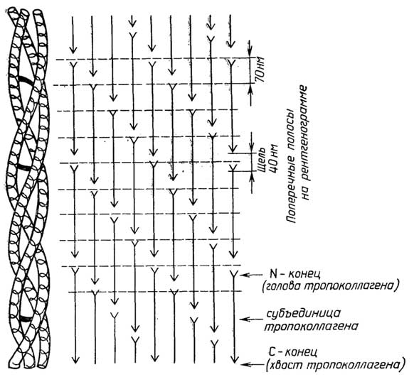
Fig. II.33. Structure of collagen.
Right: arrangement of tropocollagen subunits within fibrils; left: structure of the three-stranded collagen helix subunit with covalent cross-links between them.
Collagen fibers are virtually non-stretchable. This is due, firstly, to the structure of the collagen helix itself, which is already sufficiently stretched and rigid owing to the presence of a significant amount of proline and its derivatives, and secondly, to the presence of a large number of covalent cross-links and non-covalent interactions both within and between the collagen fibers.
Elastins
Elastins are the primary component of ligaments connecting bones. They form the basis of blood vessel
walls and serve as the second protein component of connective tissue.
The secondary structure of elastins is a unique helix characteristic exclusively of this protein. It forms globular subunits called tropoelastin or α-elastin, with a diameter of 3 nm, a molecular weight of 74,000, and approximately 800 amino acid residues. The exact folding of the helix within the globule remains unclear. However, it is known that the polypeptide chains are dominated by hydrophobic

Fig. II.34. Two types of covalent lysine cross-links in collagen and elastin.
amino acid residues, predominantly aliphatic ones. Preliminary data suggest that the helix inside the globule is arranged such that these hydrophobic side groups face inward. The native structure of elastins (Fig. II.35) is formed from tropoelastin globules cross-linked into a single network by both lysine bonds (see Fig. II.34) and specialized bonds based on desmosine and isodesmosine (see Fig. II.2), which are found exclusively in elastins. As in collagens, cross-links are absent in embryonic elastins, sparse in young organisms, but increase rapidly in number with age.
The distinctive feature of elastins lies in their ability to stretch several-fold in all directions and return to their original state upon load removal, while

Fig. II.35. Structure of elastin:
A – lysine cross-links; Б – desmosine and isodesmosine cross-links; В – tropoelastin globules.
maintaining high tensile strength. This property is explained by the fact that stretching disrupts the hydrophobic interactions within tropoelastins and alters their microenvironment, though this process is fully reversible. The high tensile strength in the stretched state is attributed to the presence of covalent cross-links.
Myosins
Myosins are the major proteins of muscle tissue (accounting for up to 60% of total protein content).

|
Fig. II.36.Schematic diagram of a myosin molecule: |
Fig. II.37.Diagram of the structure of a thick filament in vertebrate muscle tissue: |

The myosin molecule from striated muscle (m. w. 470,000) with a length of 150 nm has a complex shape (Fig. II.36). It consists of a long rod, at one end of
which are two somewhat elongated globules exhibiting enzymatic activity toward adenosine triphosphate (ATP). The molecule is composed of two identical heavy chains and two pairs of light chains. The heavy chains (m. w. 200,000 each) have an α-helical conformation and are twisted into a supercoil, forming the backbone of the molecule and terminating in the end globules. These are among the longest known polypeptide chains, containing approximately 1,800 amino acid residues each. Each terminal globule contains two additional light chains (m. w. 18,000 each) folded into their own globular domains, which are non-covalently linked to the main chain. Treatment with enzymes that cleave polypeptide chains at specific sites (see Fig. II.36) yields subfragments. Trypsin splits the molecule into light meromyosin (LMM) and heavy meromyosin (HMM). Upon treatment with papain or prolonged digestion with trypsin, the latter yields two S1 subfragments and an S2 subfragment. Although the myosin molecule is generally rigid, flexible hinge regions are located at the cleavage sites. It is quite appropriate to consider these hinge domains as connecting four distinct regions within the molecule: two S1 domains, S2, and LMM.
As a result of the non-covalent interaction of 300–400 myosin molecules, a thick filament of muscle tissue is formed, which has a diameter of 16 nm and a length of 1,500 nm. Its cross-section contains two to three dozen molecules. The packing of myosin molecules within the thick filaments is such (Fig. II.37) that they join "tail-to-tail" in the middle, forming a 150 nm central zone, whereas toward both ends of the thick filament they assemble "head-to-tail," with the HMM portions of the molecules protruding outward. Furthermore, the molecular packing is staggered, similar to that in collagen. As a result, the protruding regions are arranged in a helical pattern along the thick filament. The tail-to-tail connection in the bare zone lacking globular heads is mediated by the so-called M-protein.
Key features of myosin:
the molecule consists of several rigid segments connected by flexible hinges;
enzymatic activity is localized in the globular head (S1 subfragments);
the polypeptide chains of the head contain cysteine residues (SH-groups).
Tropomyosins
Tropomyosins are fibrillar proteins of muscle tissue (constituting about 5% of total protein content). By analogy with collagen and elastin, one might assume that tropomyosin is a subfragment of myosin, but this is incorrect. Tropomyosin is an independent protein with its own specific function, yet its molecular
structure closely resembles that of the myosin molecule, or more precisely, its tail region.
The tropomyosin molecule is relatively rigid and rod-shaped, with a length of 41 nm and a diameter of 2 nm. It is a supercoil composed of two very similar

Fig. II.38.Diagram of the structure of a thin filament in vertebrate muscle tissue. Each tropomyosin strand lies nearly within the groove between the F-actin strands, such that it contacts only one of them. Each tropomyosin molecule spans the length of seven G-actin subunits. The troponin protein, consisting of three globular subunits and playing a crucial regulatory role, is located at the junction points of adjacent tropomyosin molecules.
polypeptide chains, each having an α-helical conformation and a molecular weight of 70,000. Two strands of end-to-end joined tropomyosin molecules, wound together with the F-actin double helix, form the thin filament of muscle tissue (Fig. II.38).
Fibrins
Fibrins are proteins of a blood clot (thrombus).
The fibrin molecule (m. w. 340,000) consists of six polypeptide chains of three types linked together by disulfide bridges. Its subunit formula is (αβγ)2. It has a rod-like shape with two terminal globular domains and one central globular domain (Fig. II.39). The exact folding of the polypeptide chains within the molecule remains unknown. Oligosaccharides are attached via asparagine residues at certain sites. Under physiological conditions, fibrin molecules aggregate to form extended fibrils, in which they pack in parallel arrays staggered by half a molecule (Fig. II.39). These fibrils, oriented in various directions, give rise to a soft clot. It consists of soluble fibrin, since the formed fibrillar clot can be redissolved into monomers by shifting the pH of the medium into the acidic (<4.5) or alkaline (>9) range. The soft clot fibrin is also soluble in 1 M urea. However, after some time, this solubilization becomes impossible because the soft clot transforms into a hard clot. This conversion involves the enzymatic formation of covalent cross-links (Fig. II.39, C) between the side chains of glutamine and lysine residues in adjacent molecules.

Fig. II.39.Structure of fibrin:
A - shape of the fibrin molecule; B - packing of monomers within the fibril, showing transverse covalent cross-links within the fibril and potential cross-links with neighboring fibrils during hard clot formation; C - formation of a covalent cross-link
§ 7.
General remarks on the structure
and properties of fibrillar proteins
High tensile strength and the insolubility of many of these proteins in water are primarily explained by their structure—specifically, the presence of covalent bonds that cross-link both the polypeptide chains within the fibrils (collagen) and the fibrils themselves into a single functional network (α-keratin, collagen and elastin of adult animals, and the fibrin of a hard blood clot).
Fibrillar proteins that lack covalent cross-links can be dissolved in:
salt solutions (muscle myosin, embryonic or newborn animal collagen and elastin);
hot water (tropomyosin);
altered pH environments (soft blood clot fibrin);
solutions containing urea (soft blood clot fibrin).
Fibrillar proteins, which are actually polymers of globular proteins, are readily soluble even in distilled water (actin F).
The capacity of some of these proteins for reversible stretching is due to the presence of helical structures in their backbone that are not cross-linked internally by covalent bonds, but are held together solely by weak forces, such as hydrogen bonds (α-keratin) or hydrophobic interactions (elastin).
1α-Helix can also be designated as a 3.613-helix. It is currently recommended to designate helices by the integer number of residues contained within an integer number of turns. For example, the α-helix, 310-helix, and π-helix should properly be referred to as the 185-helix, 31-helix, and 225-helix, respectively.
2In α-, 310-, and π-helices, hydrogen bonds are formed between the end (NH group) of the n-th residue and the start (CO group) of the corresponding residue (further along the chain).
3In humans, additionally, the following have been discovered: hemoglobin A2(α2δ2)—2% of the total amount; hemoglobin α2ε2—in human embryos; hemoglobin F(α2γ2)—human fetal hemoglobin, as well as other hemoglobins in small quantities. As can be seen, α protomers are common to all human hemoglobins. The β, δ, γ, ε protomers show little difference in their primary structure, and their tertiary structures are practically identical.
Chapter 9
FUNCTIONAL DIVERSITY OF PROTEINS
AND THEIR CHARACTERISTICS
§ 1.
Functional
Classification of Proteins
According to the functions they perform, proteins are generally divided into several classes.
Enzymes are the most numerous group (numbering about 2,000). Typically, they are globular proteins, though fibrillar ones are also known (myosin). They possess catalytic activity, facilitating the chemical reactions that occur in living organisms.
Hormones are endogenous regulatory proteins of the organism, such as insulin (which regulates glucose metabolism), somatotropin (growth hormone), and others, which are capable of influencing the course and direction of metabolic processes in the body in extremely small quantities.
Toxins are exogenous proteins, such as cholera and diphtheria toxins, botulinum toxin (a metabolic product of the anaerobic bacterium Clostridiym botutinum), ricin (from castor bean seeds), and snake venoms. Like hormones, they profoundly affect metabolic processes even at very low concentrations.
Structural proteins form part of virus coats, cell walls, and skin. They are the major component of the connective tissue in tendons, cartilage, ligaments, hair, wool, turtle shells, and serve as the framework of bones. Thanks to them, the organism is held together as an integrated whole.
Transport proteins carry molecules and ions either between organs or across cell membranes. For example, hemoglobin transports O2, CO2, and H+; serum albumin transports fatty acids; and β-lipoproteins transport lipids.
Storage proteins include seed proteins (gliadin in wheat, zein in maize), ovalbumin, and milk casein. They are essential for the development of the organism in its early stages.
Motor proteins—such as muscle proteins (myosin and actin), as well as tubulin in cilia and flagella—provide the capacity for movement to the organism and/or its parts.
Protective proteins safeguard the organism against external destruction or promote its survival upon injury. Here are a few examples:
toxins produced by plants protect them from being eaten by animals, whereas toxins from insects (such as bees) or certain vertebrates (such as toads and venomous fish) defend them against predators;
structural proteins, as components of virus coats, cells, and organisms (i.e., skin, hair, shells), also perform a protective function against adverse environmental factors or organisms residing in the environment;
upon injury to blood vessels, a protective system of blood clotting and clot formation is triggered, a process involving a large group of proteins;
in response to the invasion of foreign proteins, other macromolecular compounds, viruses, or cells—collectively termed antigens—higher vertebrates are capable of synthesizing, within several days, specialized immune defense proteins called antibodies, which bind to antigens with high specificity and neutralize them;
in addition to antibodies, certain cells of higher vertebrates, responding to the entry of a specific type of virus into the body, can synthesize within a day or two specialized proteins called interferons, which bind to healthy cells and protect them from infection by the same or any other type of virus.
Proteins with special functions include, for example, receptor proteins that ensure the transmission of impulses between nerve cells or the perception of light; proteins that protect the blood of fish inhabiting cold seas from freezing, and others.
§ 2.
Species Specificity —
a Characteristic Feature of Proteins
Homologous proteins—that is, proteins performing identical functions in organisms belonging to different species—differ immunologically. This means that when such proteins are introduced into the bloodstream of a vertebrate animal (or shark), they induce the formation of antibodies with distinct structures, with the degree of difference increasing as the evolutionary relationship between the species becomes more distant.
Homologous proteins from different organisms of the same species may also exhibit immunological differences.
Section B
CARBOHYDRATES AND CELL WALLS
§ 1.
General Characteristics, Functions,
and Classification of Carbohydrates
Carbohydrates are polyhydroxy aldehydes (aldoses) and polyhydroxy ketones (ketoses), as well as their derivatives. The name "carbohydrates" stems from the fact that the composition of many of them can be expressed by the formula (CH2O)n, where n ≥ 3. Carbohydrates are the primary dry-mass components of plant cells, but their functional significance is also immense in bacterial and animal cells.
The functions of carbohydrates can be divided into three main groups:
functions related to their ability to act as carriers and stores of both chemical energy and carbon atoms; the oxidation of 1 g of carbohydrates can release about 4 kcal of energy, which is utilized in various metabolic processes, while carbon atoms are essential for the organism to biosynthesize not only carbohydrates themselves, but also proteins, nucleic acids, and lipids;
functions associated with the structural role of carbohydrates; as vital components of bacterial and plant cell walls, as well as animal cell coats, they perform not only a supportive function but also many others simultaneously. For instance, they serve as antigenic determinants of cells and numerous compounds—meaning that the presence of carbohydrate tags on the surface of cells or macromolecules allows the organism to distinguish its own cells and macromolecules from foreign ones. Carbohydrate components on the protein surface frequently protect the latter from the destructive action of the external environment or enzymes. Furthermore, carbohydrate components of cell walls and coats also act as receptors for the specific binding of various metabolic regulators, such as hormones and toxins, as well as neurotransmitters during nerve impulse transmission;
other functions: they prevent blood clotting and, in some fish, protect against freezing; they impart a mucous consistency to substances coating the epithelial tissues of the respiratory and digestive tracts, and act as lubricants in joints and other areas; they function as antibiotics and various physiologically active substances. Vitamin C is also classified as a carbohydrate.
The classification of carbohydrates is based on their division into simple and complex sugars.
Simple sugars are monosaccharides and their derivatives, while complex sugars are oligosaccharides and polysaccharides along with their derivatives.
Monosaccharides are aldoses and ketoses whose molecules contain from 3 to 7 carbon atoms. Sometimes, aldoses and ketoses with 8 and 9 carbon atoms, referred to as higher sugars, are also included in this group.
Oligosaccharides can be viewed as the product of polycondensation of 2 to 10 monosaccharide molecules or their derivatives.
Polysaccharides (glycans) result from the polycondensation of a large number of monosaccharide molecules or their derivatives. Among them, a distinction is made between homopolysaccharides, consisting of residues of a single monosaccharide, and heteropolysaccharides, which contain alternating residues of different monosaccharides (usually no more than four), such that the repeating unit is the residue of a small oligosaccharide. The molecular weight of glycans can reach hundreds of millions.
Chapter 10
MONOSACCHARIDES AND OLIGOSACCHARIDES –
SOLUBLE SUGARS
§ 1.
Monosaccharides
Monosaccharides are classified according to the number of carbon atoms in their molecules into trioses, tetroses, pentoses, hexoses, etc. Instead of the prefix "keto-", the suffix "-ulose" can be used; for example, "pentulose" instead of "ketopentose". The numbering of carbon atoms begins from the end closest to the aldehyde or carbonyl (keto) group. For the most common monosaccharides, trivial names are widely used, such as fructose. The simplest monosaccharides are trioses:

Figure II.40 shows the most common aldoses and ketoses found in nature. Monosaccharides are depicted using Fischer projection formulas.
Stereoisomerism is a property characteristic of all monosaccharides except dihydroxyacetone. The universally accepted system for carbohydrates is the

Fig. II.40. Most common hexoses and pentoses
DL-system of stereoisomerism, which is based on the conformations of D- and L-glyceraldehyde. The assignment of a monosaccharide to the D- or L-series is determined by the configuration of the hydroxyl group at the chiral (asymmetric) carbon atom with the highest locant number. This configuration is a property of the molecular structure and is unrelated to the ability of a solution of that monosaccharide form to rotate the plane of plane-polarized light in either direction (i.e., to the right or left).
Enantiomers are D- and L-stereoisomers.
Diastereomers are stereoisomers that are not enantiomers; for example, D-glucose is a diastereomer with respect to any aldohexose (in either the D- or L-form) except L-glucose.
Epimers are pairs of diastereomers that differ in the configuration of the hydroxyl groups at a specific carbon atom; for example, D-glucose and D-mannose are epimers at the C2 atom, and D-glucose and D-galactose are epimers at the C4 atom.
Cyclic structures of monosaccharides represent the primary forms in which pentoses, hexoses, and heptoses exist in aqueous solutions.
Anomers are pairs of stereoisomers designated as α- and β-, the formation of which is associated with the existence of monosaccharides in the cyclic form of intramolecular hemiacetals or hemiketals. The phenomenon of interconversion of two anomers via a transient linear form (aldehyde or ketone) is termed mutarotation. This process is manifested by a change in the angle of rotation of the plane of polarization of plane-polarized light when the latter passes through solutions of anomers. Mutarotation is accelerated by an increase in the pH of the medium.
The cyclic structures of monosaccharides are conventionally represented by Haworth projection formulas. The cyclization of pentoses, hexoses, and others typically results in the formation of either a pyranose form, i.e., a six-membered ring resembling the structure of pyran (Fig. II.41), or a furanose form, i.e., a five-membered ring resembling the structure of furan (Fig. II.42). The pyranose forms of pentoses and hexoses are generally more stable in solutions, whereas their furanose forms occur as constituents of oligosaccharides.
Conformational formulas, unlike Haworth formulas, provide insight into the spatial arrangement of the atoms within the ring.
The pyranose form of pentoses and hexoses exhibits three conformations (Fig. II.43): "chair", "boat", and "skew-boat" ("twist"). The most stable is the "chair" conformation, which in turn exists in two conformational variants: "C1" and "1C" (Fig. II.44). The former is characteristic of D-series monosaccharides, while the latter is typical of the L-series. The "boat" and "skew-boat" structures are more flexible and possess several conformational variants. The transition from "chair" to "boat" involves an energy change of 5–6 kcal · mol.
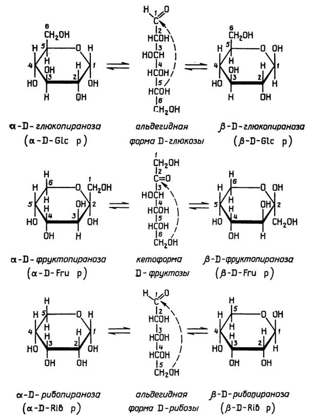
Fig. II.41. Cyclization of D-glucose, D-fructose, and D-ribose yielding pyranose anomers depicted by Haworth formulas
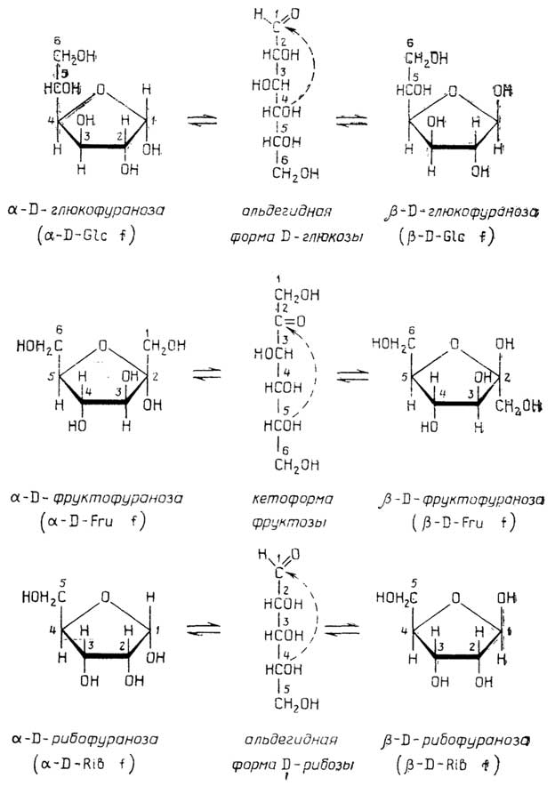
Fig. II.42. Cyclization of D-glucose, D-fructose, and D-ribose yielding furanose anomers depicted by Haworth formulas

Fig. II.43. Conformational forms of the pyranose ring of pentoses and hexoses
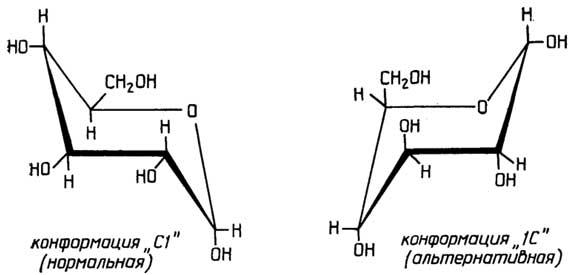
Fig. II.44. Variants of the "chair" conformation
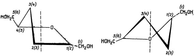
Fig. II.45. Conformational forms of the furanose ring of pentoses and hexoses
The furanose structure of pentoses and hexoses can exist in either the "envelope" or the "skew-boat" ("twist") conformation (Fig. II.45). The "envelope" conformation is more stable. Both conformations have several variants, which is associated with

Fig. II.46. Variants of the "envelope" (A–G) and "skew-boat" (D, E) conformations
the different spatial orientations of the C2 and C3 atoms relative to the C5 atom (Fig. II.46).
A significant effect on conformational stability is exerted by the spatial arrangement of substituents at the carbon atoms of the ring. The equatorial
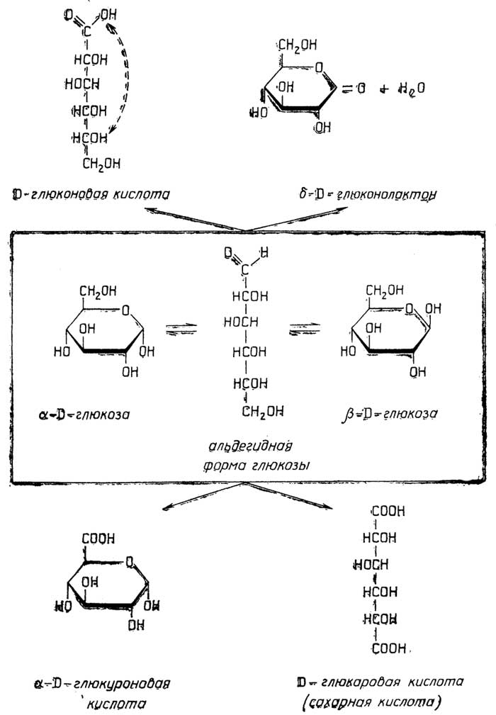
Fig. II.47. Sugar acids: oxidation products of D-glucose
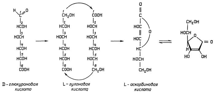
Fig. II.48. L-Ascorbic acid (vitamin C)
arrangement is preferred over the axial one. In β-glucose, substituents at all atoms are arranged equatorially, whereas in α-glucose, the hydroxyl group of the anomeric C1 atom is positioned axially and the remaining substituents are equatorial.
§ 2.
Biologically Important
Monosaccharide Derivatives
Sugar acids are produced by the oxidation of monosaccharides at the aldehyde group or the C6 hydroxyl group. Depending on which groups are oxidized, aldonic, uronic, and aldaric acids are formed. Thus, upon the oxidation of glucose

Fig. II.49. Sugar alcohols
(Fig. II.47), these are D-gluconic acid (more stable in the δ-lactone form), D-glucuronic acid, and D-glucaric (saccharic) acid. The first of these plays an important role in the metabolism of living organisms as an intermediate, the second

Fig. II.50. Monosaccharide esters: phosphate and sulfate (top) and glycosides (bottom)
is a component of many biologically vital compounds, and the third occurs rarely.
Ascorbic acid (vitamin C) belongs to the sugar acids. In the bodies of animals—with the exception of primates, guinea pigs, and humans—it is synthesized from glucose via D-glucuronic acid (Fig. II.48).
Sugar alcohols are products of monosaccharide reduction at the aldehyde or ketone group. The best known are sorbitol, dulcitol, mannitol, ribitol, xylitol, and myo-inositol (shown in Fig. II.49).
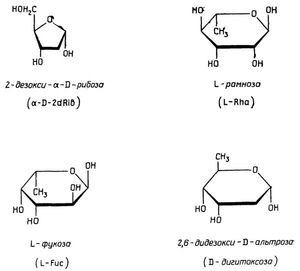
Fig. II.51. The most well-known deoxy sugars
Monosaccharide esters can be formed in two ways: either as esters or as ethers. In the first case, they are products of the reaction between the hydroxyl groups of monosaccharides and acids such as phosphoric, sulfuric, or acetic acid, e.g., α-D-glucose-6-phosphate or β-D-glucose-2-sulfate (Fig. II.50). In the second case, they are products of the reaction between the hydroxyl groups of monosaccharides and the hydroxyl groups of various natural compounds, such as alcohols and phenols. The non-sugar moiety of such esters is called an aglycone, the esters themselves are termed glycosides (Fig. II.50), and the bond that links them is referred to as an -O-glycosidic bond.
Deoxysugars (Fig. II.51) play a crucial role as components of cell surface antigenic determinants and are part of many biologically important compounds and antibiotics; 2-deoxy-D-ribose is a component of DNA.
142
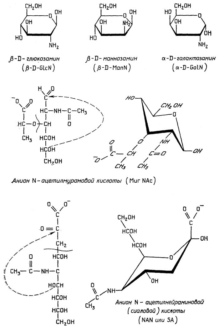
Fig. II.52.
Most common aminosugars
and their derivatives
Aminosugars and their derivatives (Fig. II.52) are structural components of many natural glycans. They are particularly abundant in polysaccharides that perform structural functions.
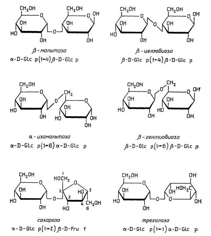
Fig. II.53.Most common disaccharides
§ 4.
Oligosaccharides
Depending on the number of monosaccharide residues, oligosaccharides are classified as disaccharides, trisaccharides, tetrasaccharides, etc. Based on the presence or absence of an anomeric carbon atom, they are classified as reducing or non-reducing, respectively. The most common disaccharides, along with their names and accepted designations, are shown in Fig. II.53. Trisaccharides (raffinose—a compound of galactose, glucose, and fructose) are less common. Disaccharides and trisaccharides typically serve as
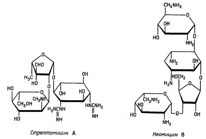
Fig. II.54.
Antibiotic carbohydrates
transport or storage forms of sugars. Higher-order oligosaccharides in their free form are relatively rare. In plants and certain insects, for example, they function as antifreeze agents. More commonly, higher-order oligosaccharides occur in conjugates with proteins (glycoproteins) or lipids (glycolipids). Certain antibiotics also belong to oligosaccharides and their derivatives (Fig. II.54).
Chapter 11
POLYSACCHARIDES (GLYCANS) —
THE MAJOR CARBOHYDRATES
OF LIVING NATURE
The chains of these natural polymers differ from those of other natural polymers, such as proteins and nucleic acids, in that they can be highly branched.
§ 5.
Storage polysaccharides
Storage polysaccharides are invariably homopolysaccharides and almost exclusively intracellular substances.
Starch is the primary storage polysaccharide in plants, deposited within cells in the form of granules. It occurs in two forms: α-amylose and amylopectin, which are found in plants either separately or as mixtures.
α-Amylose (with a molecular weight ranging from several thousand to 500,000) consists of linear chains comprising hundreds or even thousands of α-D-glucopyranose molecules linked by α(1→4) glycosidic bonds. Amylopectin differs from α-amylose in that its chains are extensively branched, with branch points occurring on average every 20–30 residues. These branches are formed by additional α(1→6) glycosidic bonds (Fig. II.55), making the amylopectin molecule a highly branched structure in which only a single branch possesses an α-D-glucose residue with a reducing anomeric C1 atom. The molecular weight of amylopectin reaches tens of millions.
Glycogen is the storage polysaccharide of animal cells, though it is also found in fungi and even certain plants. In animals, it is typically located in liver and muscle cells. Its structure is analogous to that of amylopectin, with the exception that branching is more frequent, occurring every 8–12 α-D-glucose residues. The molecular weight of glycogen can reach up to 100 million.
Dextrins are remnants of amylopectin or glycogen representing their branched "cores" left over after the removal of all linear outer branches through the selective hydrolysis of α(1→4) bonds, starting from the branch ends inward to the branch points. Thus, dextrins are artificially derived products.
Dextrans are storage polysaccharides found in bacteria and yeasts. They are poly-D-glucopyranoses whose main chains are formed by α(1→6) bonds, while numerous branches are formed by α(1→2), α(1→3), and α(1→4) bonds. The molecular weight of dextrans is in the hundreds of millions. High-molecular-weight dextrans, much like proteins, exhibit antigenic properties.
Other storage polysaccharides (see Fig. II.55), which are less common, also occur in living nature. Examples include poly-β-D-fructofuranose (artichoke inulin), poly-α-D-galactopyranose (found in certain plants), and poly-β-D-galactofuranose (found in certain fungi).

Fig. II.55.Storage polysaccharides
§ 6.
Structural polysaccharides
They can be conventionally divided into primary and secondary (or auxiliary) types. The former serve as the main "reinforcing" material in cell walls (of plants, algae, fungi, and even a single lower animal species) or in extracellular structures, such as the exoskeleton of many arthropods. The latter perform an auxiliary role by "cementing" the primary structural framework into a cohesive whole.
Primary structural polysaccharides
Cellulose is common to all plants, but is also found in bacteria, fungi, and lower animals (tunicates). It is a linear homopolysaccharide composed of β-D-glucopyranose units linked by β (1→4) bonds (Fig. II.56). The number of D-glucose residues in the polymer chain ranges from several thousand up to 10,000, with a molecular weight apparently reaching 1–2 million.
Chitin is widely distributed in the animal kingdom and is also abundant in fungi. It is a homopolysaccharide of N-acetylglucosamine whose molecules form linear chains analogous to those of cellulose (Fig. II.56). In nature, chitin is invariably bound to proteins, lipids, and other components, making its isolation in pure form quite difficult. Chitin is precisely what provides the rigidity of the exoskeleton in many arthropods.
Other major structural polysaccharides are found in algae and fungi. Most frequently, these are homopolysaccharides of D-mannose, D-galactose, and D-xylose, which can form either branched or linear chains. Examples of both types are illustrated in Fig. II.56.
Auxiliary Plant
Structural Polysaccharides
Hemicellulose is a collective term for various highly branched polysaccharides (Fig. II.57) consisting of xylan, araban, galactan, and mannan chains, where each chain may additionally incorporate D-xylose, D-glucose, D-galactose, D-mannose, L-arabinose, L-fucose, and sometimes glucuronic acid derivatives after a specific number of units.
Pectins are much less branched polysaccharides compared to hemicellulose (Fig. II.57). They exist as protopectin and true (soluble) pectin. Protopectin not only contributes to cell wall structure but is also the major component of the intercellular substance, particularly in fruits and vegetables. Its chains consist of poly-D-galacturonic acid, whose carboxyl groups are esterified with methanol to varying degrees. The chains also include a certain number of
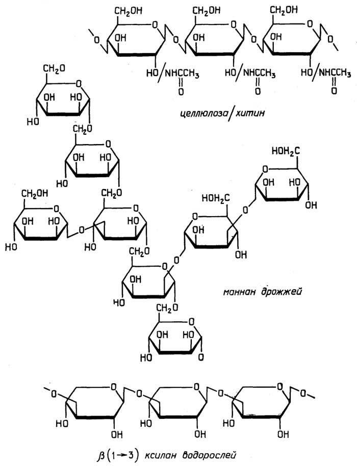
Fig. II.56. Major structural polysaccharides

Fig. II.57. Auxiliary plant structural polysaccharides
residues of other monosaccharides, such as D-galactose, L-arabinose, and L-rhamnose. The chains typically bear branches composed of D-galactose and L-arabinose, through which protopectins link with other polysaccharides, both major and auxiliary. The cleavage of these bonds (in a mildly acidic environment) converts protopectin into soluble pectin, which is typically present in the juice of fruits and vegetables.
Auxiliary Algal
Structural Polysaccharides
Because algal cell walls experience significantly lower mechanical stress compared to land plant cell walls, the role and proportion of major structural polysaccharides in algae are negligible. Auxiliary
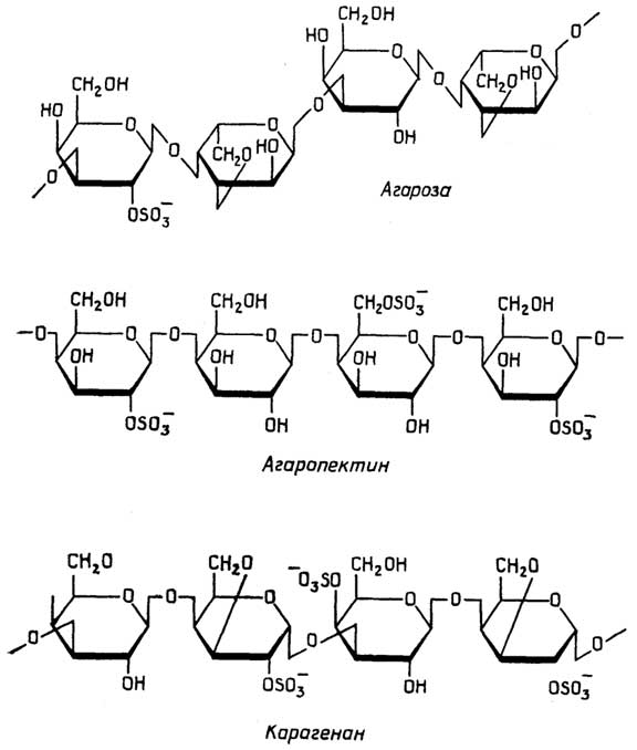
Fig. II.58. Auxiliary algal polysaccharides
structural polysaccharides in algae serve as the basis of the intercellular matrix.
Agar-agar (Fig. II.58) consists of two components, agarose and agaropectin, and is extracted from red seaweeds. Agarose is formed from chains whose repeating unit is a disaccharide of β-D-galactose and 3,6-anhydro-α-L-galactose. The intersugar bond within the disaccharide is β (1→4), and the linkage between disaccharides is α (1→3). Some of the hydroxyl groups may be esterified with sulfuric acid. Agaropectin is composed of poly-β-D-galactose chains in which the hydroxyl groups are partially esterified with sulfuric acid.
Carrageenan (Fig. II.58), like agar-agar, is extracted from red algae and bears a strong resemblance to it. Its chains are built from a disaccharide of β-D-galactose and 3,6-anhydro-α-D-galactose. The bond within the disaccharide is β (1→4), and the linkage between disaccharides is α (1→3). The hydroxyl groups are partially esterified with sulfuric acid.
Algin, found in brown and other marine algae, is an acidic homopolysaccharide of D-mannuronic acid.
§ 7.
Polysaccharides
with Diverse Functions
This group encompasses all polysaccharides that do not classify as storage or structural. They function extracellularly, either associated with the cell surface or in a free state as extracellular substances.
Glycosaminoglycans
Glycosaminoglycans (mucopolysaccharides) are a group of heteropolysaccharides sharing the common feature of containing amino sugar residues alongside acidic groups—either carboxyl or sulfate groups. They form the foundation of the intercellular matrix in animal tissues, particularly connective tissue. Most of them are typically bound to proteins.
Hyaluronic acid (Fig. II.59) is the most widespread glycosaminoglycan. This heteropolysaccharide consists of linear chains whose repeating unit is a disaccharide of β-D-N-acetylglucosamine and β-D-glucuronic acid. The linkage within the disaccharide is β (1→4), and the bond between disaccharides is β (1→3). Hyaluronic acid occurs in both bound and free forms. It is especially abundant in the synovial fluid of joint capsules, where its viscous solutions act as a protective lubricant. In many bacteria, it forms part of the cell envelope.
Chondroitin sulfate, dermatan sulfate, and keratan sulfate are similar to hyaluronic acid in their structure (Fig. II.59), tissue localization, and

Fig. II.59. Glycosaminoglycans: extracellular polysaccharides of animal tissues
functions, but they are found exclusively in protein-bound forms, particularly in mucins (see p. 159, "Proteoglycans").

Fig. II.60. Heparin and heparan sulfate
Heparin (Fig. II.60) is structurally similar to many mucopolysaccharides, but serves different functions. Bound to a protein, it is found on the surface of many cells, particularly blood cells, where it acts as an anticoagulant. Heparin is synthesized and stored in mast cells. Heparan sulfate differs from heparin only in having fewer sulfate groups and more N-acetyl groups.
Gums
These are sticky substances secreted by plants at sites of damage, and they are also acidic heteropolysaccharides. Their chains consist of D-galactose and D-glucuronic acid residues, and also include a certain amount of L-arabinose and L-rhamnose.
§ 8.
Levels of Structural
Organization of Polysaccharides
The concept of the "level of structural organization"—i.e., primary, secondary, tertiary, and quaternary structures—is fully applicable to polymers such as polysaccharides.
Primary Structure
The primary structure of polysaccharides is formed through the polycondensation of one or several (usually no more than four) different monosaccharides. It can be linear or branched to varying degrees. Linear chains are characteristic of principal structural glycans (cellulose, chitin, and the D-xylan of certain algae), as well as accessory structural glycans of animal tissues (glycosaminoglycans). Branched chains are typical of storage glycans (amylopectin4, glycogen, dextran) and accessory structural glycans of plant tissues (hemicellulose, pectin).
The difference between the primary structure of polysaccharides and that of proteins is that:
polysaccharide chains consist of repeating units, whereas protein chains typically do not contain repeats; those proteins (such as collagen, extensin) whose chains consist of a small set of amino acids with repeating units bear a certain resemblance to some glycans (e.g., algal D-xylan);
unlike protein chains, polysaccharide chains can be branched.
Secondary Structure
The secondary structure of polysaccharides and proteins arises due to similar underlying principles. In protein chains, one rigid peptide bond alternates with two flexible single bonds. In glycan chains, the role of the rigid bond is played by the monosaccharide ring, followed by the glycosidic bond, which consists of two flexible single bonds through which the oxygen atom links the monosaccharide residues (Fig. II.61). Thus,
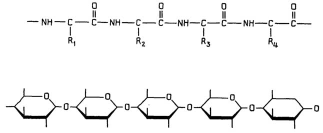
Fig. II.61.Comparison of the flexibility of protein and polysaccharide chains
it can be considered that during the formation of secondary structure, the flexibility of the polymer chain in both glycans and proteins is determined by the same number of degrees of freedom. This is confirmed in practice: the secondary structures of glycans strongly resemble those of proteins.
Ribbon-like structures, similar to protein β-sheets, are characteristic of cellulose and chitin. Their formation is explained, firstly, by the fact that cellulose and chitin tend to form rigid linear chains, a rigidity caused by the equatorial orientation of the hydroxyl groups of β-D-glucose and the resulting intra-chain hydrogen bonds (Fig. II.62), and secondly,

Fig. II.62.Formation of intra-chain hydrogen bonds in poly-β-D-glucose
by the fact that the parallel arrangement of such rigid chains is stabilized by the formation of numerous inter-chain hydrogen bonds (Fig. II.63). These same factors
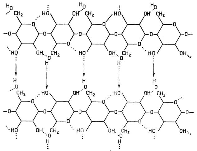
Fig. II.63.Secondary structure of cellulose resembling a parallel protein β-sheet
account for the poor solubility of cellulose (β-proteins are also poorly soluble).
Helical structures are formed by many glycans:
a left-handed single helix with 6 monosaccharide residues per turn is characteristic of α-amylose;
a left-handed single helix with 4 residues per turn is characteristic of hyaluronic acid;
a left-handed double helix with 3 residues per turn in each chain is found in agarose;
a left-handed double helix with parallel or antiparallel chain orientation and 6 residues per turn for each chain is apparently also possible for α-amylose (and for the linear regions of amylopectin);
a right-handed double helix with 3 residues per turn in each chain forms the basis of the carrageenan structure. Its unique feature is that one of the chains winds around the other with a shift of half a turn.
a right-handed triple helix with parallel chain orientation and 6 residues per turn for each chain was found in D-xylan, the primary structural glycan of certain algae. It bears a certain resemblance to the collagen triple superhelix.
The main polysaccharide helical structures in cross-section are shown in Fig. II.64.
Tertiary Structure
Tertiary structure in polysaccharides refers to the spatial folding of layers and helices—or, in their absence, the spatial arrangement of polymer chains—much like in proteins. Several typical tertiary structures can be distinguished among glycans.
Rigid fibers in cellulose are formed from folded double-chain ribbons. Stabilized by numerous interchain hydrogen bonds, these fibers are quite rigid, inextensible, and capable of withstanding immense tensile loads, which is vital for plant cells. Similar fibers evidently form the basis of chitin.
Flexible fibers in the cell walls of certain algae are formed by right-handed triple helices of D-xylan, also stabilized by interchain hydrogen bonds. They closely resemble tropocollagen fibrils. Such fibers withstand substantial tensile loads while possessing significant flexibility, which is better suited for algal cells.
Complex branched compact structures of globular shape are formed by storage polysaccharides such as amylopectin, glycogen, and dextrans. α-Amylose helices, both single and in associations, can be incorporated into such globular molecules.
Complex branched loose disordered structures are characteristic of auxiliary plant structural glycans such as hemicelluloses and protopectins. This architecture is dictated by their function: to "glue" the fibers of the primary structural polysaccharides into an intact cell wall, and the cells into a tissue.
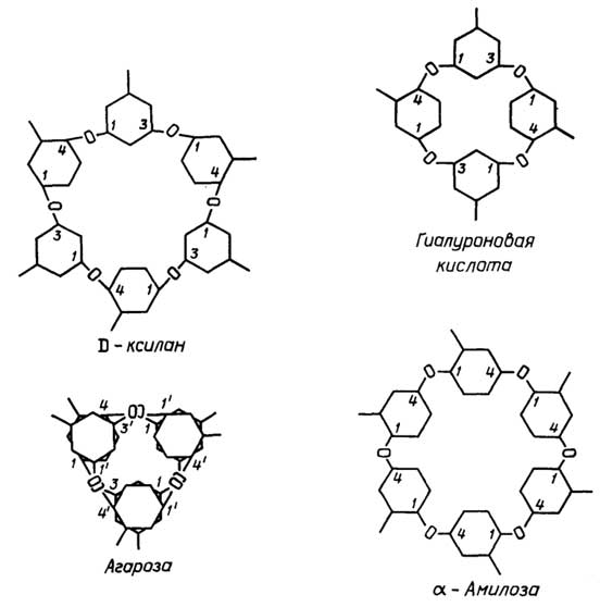
Fig. II.64. Cross-sectional views of polysaccharide helical structures
Spatially reticular loose structures, whose threads consist of multiple helices, are characteristic of gel-forming acidic polysaccharides such as agar-agar, carrageenan, algin, soluble pectins, and gums. These structures are very similar to the previous ones, differing only in the presence of a large number of helices. It can be said that they "glue" together not the primary structural glycans, but rather a specific amount of water along with dissolved substances, resulting in semi-rigid structures.
Spatially disordered mobile structures can be attributed to hyaluronic acid. They are characterized by the easy disruption and reformation of bonds between adjacent helices; therefore, hyaluronic acid gels function not merely as an intercellular substance, but also as an extracellular lubricant wherever tissue friction occurs.
Quaternary Structure
Quaternary structure in polysaccharides is simply the next stage of spatial organization following the tertiary structure. Manifestations of quaternary structure undoubtedly include:
the formation of crystalline regions within the cellulose structure, consisting of orderly arranged cross-layered cellulose fibers;
the formation of crystalline regions in starch granules, apparently consisting of orderly and densely packed amylopectin globules that incorporate the α-amylose helix; the latter, albeit in the form of more twisted double helices with parallel or antiparallel chain orientation, appears also capable of forming crystalline regions within starch granules.
4α-Amylose can be considered an intermediate product in the formation of amylopectin.
Chapter 12
CARBOHYDRATE COMPOUNDS
WITH OTHER SUBSTANCES
§ 9.
Molecular Complexes of Carbohydrates
with Peptides and Proteins
Peptidoglycans are structures composed of polysaccharide chains "cross-linked" by short peptides (Fig. II.65).
Proteoglycans are polysaccharide-protein compounds in which polysaccharides constitute the major part. Mucins (mucoproteins) from the mucous secretions of the digestive tract also belong to these compounds. In proteoglycans, numerous polysaccharides are attached via glycosidic bonds to an extended protein chain. It is hypothesized that such proteoglycans can aggregate into massive assemblies sharing a hyaluronic acid backbone (Fig. II.66).
Glycoproteins are complex proteins containing one or more prosthetic groups consisting of a monosaccharide, oligosaccharide, or polysaccharide (Fig. 66, B). In glycoproteins, the carbohydrate mass fraction is smaller than the protein fraction. Glycoproteins are widespread across all types of organisms, being especially abundant in animal cells. They include enzymes, antibodies, transport proteins, hormones, receptor proteins, etc.

Fig. II.65. Peptidoglycan of bacterial cell walls
The functions of the carbohydrate moiety of a glycoprotein are diverse, yet all boil down to acting as a tag that ensures the recognition of the given glycoprotein by other molecules or cells that interact specifically with it. Such
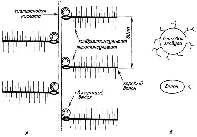
Fig. II.66. Proteoglycan (A) and glycoproteins (B)
interactions underlie the functioning of the immune system in higher animals, as well as plant defense systems against numerous diseases. A specific yet clear example of the role of carbohydrate tags is their decisive influence on blood group specificity (Fig. II.67).
The countless variations of carbohydrate tags in glycoproteins and (glycolipids) stem from a wide diversity in their composition and structure. While compositional diversity is associated with the participation of numerous isomers and their derivatives in building the carbohydrate chain, structural diversity is determined by multiple linkage patterns between monosaccharide residues with branching capabilities.
The formation of glycoproteins from proteins synthesized on the ribosomes of the rough endoplasmic reticulum occurs via enzymes located on the membranes of the Golgi apparatus. It is believed that carbohydrates primarily tag (glycosylate) proteins destined to function either as part of the cell wall or extracellularly. Proteins synthesized by the cell for its own use and which
Fig. II.67. Erythrocyte antigenic determinants determining human blood groups
must function internally (within the cytoplasm) remain unglycosylated. They are synthesized on free ribosomes not attached to endoplasmic reticulum membranes.
§ 10.
Structure of Cell Walls in Plants,
Algae, Fungi, and Bacteria
Plant cell walls, especially those of woody plants, withstand colossal loads. Their architecture resembles that of reinforced concrete. Cellulose fibers serve as the main structural material. Arranged parallel to each other, they form crisscrossing layers. Accessory structural materials include hemicellulose, protopectins, the glycoprotein extensin (whose composition and structure resemble the fibrous protein collagen), and the highly branched polymer lignin (molecular weight around 10,000), whose monomer is coniferyl alcohol (Fig. II.68).
Fig. II.68. Coniferyl alcohol—a lignin monomer. Arrows indicate potential bonding sites within the polymer |
|
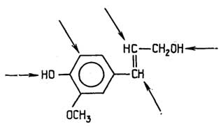
All components of cell walls are interconnected and bound with cellulose fibers into a unified structure. Lignin is found exclusively in the cell walls of woody plants, imparting the hardness that distinguishes them from herbaceous plants.
The cell walls of protists with plant-like traits, such as algae and fungi, must be sufficiently sturdy yet not as rigid as those of woody plants. Their construction principle mirrors that of plants: primary structural glycans cemented by accessory ones.
In algae, the primary structural glycans are fibers composed of cellulose and other glycans, which may form helices, such as D-xylan fibers. Poly-D-mannose fibers are also common, as well as poly-D-glucose where monomers are joined by (1→2) and (1→3) linkages. The accessory structural glycans of algae comprise various acidic gel-forming glycans prone to helix formation.
In fungi, the primary structural glycan—besides cellulose—is chitin, along with other polysaccharides, notably mannans. Accessory structural materials in fungi include glycoproteins.
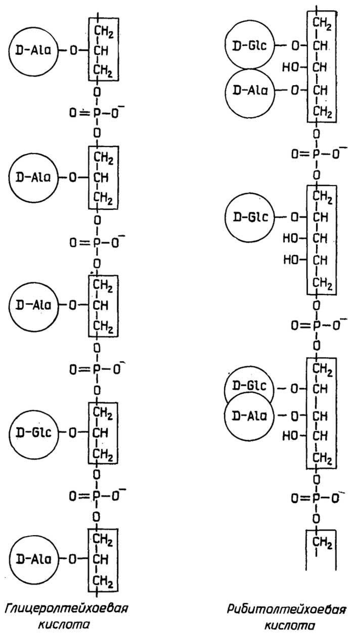
Fig. II.69. Teichoic acids
The cell walls of bacteria must be sufficiently robust to ensure microbial survival in fluctuating environmental conditions. The rigid cell walls of all bacteria except mycoplasmas are constructed from a continuous peptidoglycan, also referred to as murein. In Gram-positive bacteria, it possesses the structure depicted in Fig. II.65, with species-specific variations in peptide composition. In Gram-negative bacteria, peptide cross-links such as pentaglycine bridges are absent, and the peptide side chains of the polysaccharide chains are linked directly. Furthermore, the murein sacculus of Gram-positive bacteria is multilayered and several times thicker than that of Gram-negative bacteria. Its layers are penetrated and reinforced by teichoic acids (Fig. II.69) attached via phosphodiester bonds. The backbone of these acids consists of chains of either glycerol residues (glycerol teichoic acids) or ribitol residues (ribitol teichoic acids) joined by phosphodiester bonds. Central carbon atoms in the glycerol or ribitol residues attach amino acids (most commonly D-alanine) via ester bonds or various monosaccharides (typically D-glucose, D-galactose, their acetylamines, or L-rhamnose) via glycosidic bonds. The outer ends of teichoic acids project outward from the cell wall and determine the antigenic specificity of the microorganism. The inner ends of teichoic acids often possess a lipid character, anchoring them into the lipid plasma membrane and thus linking it to the cell wall. In addition to teichoic acids, small amounts of other polysaccharides, peptides, and proteins are found in the murein sacculus structure, which presumably perform functions similar to those of teichoic acids to a certain degree.
Section B
LIPIDS AND BIOMEMBRANES
Lipids is a collective term for highly heterogeneous substances that are sparingly soluble in water but can be extracted from living tissue by organic solvents. Unlike other classes of substances—namely proteins, nucleic acids, and polysaccharides—the majority of lipids are not polymers.
Based on general structural features and susceptibility to hydrolysis, the vast majority of lipids can be classified as either saponifiable or nonsaponifiable. Saponifiable lipids break down upon hydrolysis into several (usually no more than four) structural components. Nonsaponifiable lipids, or isoprenoids, represent either short polymeric chains of isoprene with simple organic molecules occasionally attached to one or both ends, or these compounds are isoprene derivatives.
Main functions of lipids:
storage of energy and carbon atoms (the oxidation of 1 g of neutral fats releases 9 kcal of energy);
protective function: for instance, the animal skin lipid layer protects against mechanical and temperature impacts, whereas the leaf lipid coating prevents bacterial penetration and excessive water evaporation;
major structural components of cell membranes;
metabolic regulators—these include lipid hormones, vitamins, and related compounds derived from certain fatty acids: prostaglandins and leukotrienes.
Chapter 13
MAIN TYPES OF LIPIDS
AND RELATED BIOLOGICALLY
ACTIVE COMPOUNDS
§ 1.
Saponifiable Lipids
Structural Components
The structural components of natural saponifiable lipids can be conventionally divided into five groups: A, B, C, D, E.
Components A are fatty acids and aliphatic alcohols.

Fig. II.70. Most common fatty acids
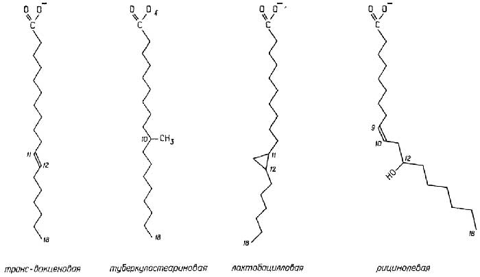
Fig. II.71. Rare fatty acids
Fatty acids of lipids are monocarboxylic acids with an aliphatic chain. About 70 of them have been discovered. Typically, they possess linear chains with an even number of carbon atoms ranging from 12 to 20. Shorter or longer chains, as well as fatty acids with an odd number of carbon atoms, are less common. Furthermore, fatty acids are occasionally found whose aliphatic chains contain substituents such as methyl,

Fig. II.72. Glycerol and sphingosine
hydroxyl, and carbonyl groups, epoxy groups, or even cyclopropane or cyclopentane rings. A significant number of lipid fatty acids, especially of plant origin, are unsaturated carboxylic acids. They may contain one or multiple double bonds, which most frequently have the cis-configuration and are extremely rarely conjugated (usually being separated by methylene groups). The melting point of fatty acids decreases with an increase in the number of double bonds in the chain and with the shortening of the chain itself. Triple bonds are also occasionally found in lipid fatty acids. The most common fatty acids are shown in Fig. II.70, while rarer ones are shown in Fig. II.71.
Aliphatic alcohols as lipid components have a chain length comparable to that of fatty acids or slightly longer (up to 32 carbon atoms). They can be saturated or unsaturated, with an even or odd number of carbon atoms.
Components B are glycerol and sphingosine (Fig. II.72). Since glycerol in lipids typically has various substituents in place of hydroxyl groups, its central carbon atom becomes chiral. In this case, natural glycerol compounds conform to L-enantiomers in accordance with the rules of stereospecific (sn) carbon atom numbering. Sphingosine differs from glycerol by the presence of a long aliphatic tail at the C3 atom and the replacement of the hydroxyl group with an amino group at the C2 atom. The aliphatic tail in sphingosine features a double bond in the trans-configuration between the C4 and C5 atoms. The most widespread is sphingosine with a chain length of 18 carbon atoms, but sphingosines with shorter (16, 17 carbon atoms) and longer chains (19, 20 carbon atoms) are also known. The amino group and the hydroxyl group at the chiral atoms C2 and C3, respectively, are in the D-position, but point in different directions in space (see Fig. II.72). Along with sphingosine, dihydrosphingosine, which lacks a double bond in the aliphatic tail, is also found in natural lipids.
Component C is orthophosphoric acid.
Components D are usually ethanolamine, choline, and L-serine, as shown in the figure:
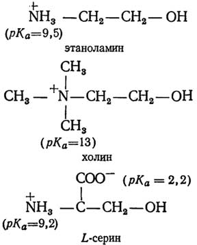
Components E are glycerol, inositol, monosaccharides, and oligosaccharides.
Classification
The classification of saponifiable lipids and their naming depend on the set of structural components and the order in which they are linked (Fig. II.73). Simple lipids consist either of two components

Fig. II.73.
Lipid classification.
Hydrophilic parts of molecules
are indicated by a dashed line
A linked by an ester bond, or of components A linked to component B, which in this case can only be glycerol. Complex lipids consist of more than two components. These include: phospholipids, having the composition ABCD; glycophospholipids — ABCE or ABCED; glycolipids with the formula ABE; sphingolipids — any lipids containing sphingosine as component B.
Simple Lipids Performing Primarily Energy-Storage and Protective Functions
Waxes are esters of fatty acids and aliphatic alcohols. They form the protective coating of leaves and fruits, as well as the protective grease of feathers and skin. They serve as the primary form of energy reserves for many microorganisms in marine plankton. The most well-known are beeswax and sperm whale spermaceti. The basis of the former is myricyl palmitate, and of the latter, cetyl palmitate:

Neutral fats (Fig. II.74) are esters of fatty acids and glycerol: mono-, di-, and triacylglycerols. The latter are the most common. Triacylglycerols may contain identical fatty acids, but more often they are different, with an unsaturated fatty acid typically attached to the C2 atom. Animal-derived triacylglycerols transition to a liquid state at higher temperatures than plant-derived triacylglycerols. The former differ from the latter by a higher content of saturated fatty acids.
Diacylglycerol ethers (Fig. II.74) differ from neutral fats in that an aliphatic alcohol, which may be saturated or unsaturated, is attached by an ether bond to glycerol at the C1 position instead of a fatty acid. They are present in living tissues in extremely small amounts.
Neutral plasmalogens (Fig. II.74) are diacylglycerol ethers with an unsaturated aliphatic alcohol, in which the double bond is located between the first and second carbon atoms and has a cis-configuration. Such 1-α,β-alkenyl-2,3-diacylglycerols, upon hydrolysis in an acidic environment, yield (in addition to two fatty acids and glycerol) an α,β-unsaturated alcohol, which immediately undergoes rearrangement into an aldehyde (plasmal).

Fig. II.74. Glycerol-based simple lipids
Some rarer lipids
with an energy storage function
Diol lipids are esters of certain diols (Fig. II.75) with components A, i.e., fatty acids and aliphatic alcohols. Both mono- and diacyl derivatives of diols are found,
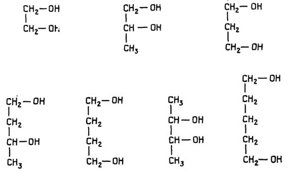
Fig. II.75. Diols as the basis of certain lipids
simple diethers, mixed alkyl, alkenyl, and acyl derivatives of diols, and other analogues of glycerol-containing lipids. Diol lipids are found in small amounts in plant and animal tissues, as well as in microorganisms. Their role apparently comes down to providing an energy reserve, and possibly a structural one as well.
Poly-β-D-hydroxybutyrate (Fig. II.76), a storage substance in many bacteria, is an "atypical" lipid. Its molecule consists of 1,500 residues of β-D-hydroxybutyric acid, the single unbranched chain of which is coiled into a tight right-handed helix. This helix, in turn, folds repeatedly to form thin (5 nm) flat sheets within the cell.
Phospholipids are the main structural components of biomembranes. These are complex lipids containing four types of components: A, B, C, D. Depending on the nature of component B, they are subdivided into glycerophospholipids and sphingophospholipids.
Glycerophospholipids are derivatives of phosphatidic acids (Fig. II.77), which are esters of glycerol with two fatty acids and orthophosphoric acid. Free phosphatidic acids are virtually undetectable in tissues, but when linked via ester bonds to ethanolamine, choline, or L-serine, they form widely distributed glycerophospholipids: phosphatidylethanolamine (cephalin), phosphatidylcholine (lecithin), and phosphatidylserine, respectively (Fig. II.77).
Glycerophospholipid ethers (Fig. II.78) with a saturated or unsaturated aliphatic alcohol attached

Fig. II.76. β-D-hydroxybutyric acid and poly-β-D-hydroxybutyrate
to the C1 atom of glycerol, occur significantly more frequently in tissues than dialkylglycerol ethers.
Phosphatidals (Fig. II.79) are plasmalogens, but unlike neutral plasmalogens, they are glycerophospholipids.

Fig. II.77. Most common glycerophospholipids
Sphingophospholipids are derivatives of ceramides (Fig. II.79), which are sphingosines (or dihydrosphingosines) acylated with a fatty acid at the amino group. Free ceramides are rarely found in tissues, but when linked through a phosphate ester bond to choline, they form widely distributed sphingolipids known as sphingomyelins.
§ 2.
Nonsaponifiable lipids (isoprenoids)
The structural building block of isoprenoids is isoprene. It acts as a monomer used to construct the oligomeric or polymeric chains of nonsaponifiable lipids. Typically, isoprene residues are joined in a periodic "head-to-tail" sequence, though "tail-to-tail" linkages also occur:

Classification
The classification of nonsaponifiable lipids is based on their traditional division into two groups: terpenes and steroids.
Terpenes
These are oligomeric and polymeric substances whose molecules are composed of isoprene residues. Terpenes whose molecules are compounds of 2, 3, 4, 6, and 8 isoprene units are referred to, respectively, as monoterpenes, sesquiterpenes, diterpenes, triterpenes, and tetraterpenes. Terpene molecules can have a linear or cyclic (even polycyclic) structure, or a structure containing both linear and cyclic regions simultaneously. Double bonds in linear regions typically have the trans-configuration, although terpenes also occur in which one or more bonds may have the cis-configuration. In terpene molecules, certain carbon atoms may bear hydroxyl, carbonyl, and carboxyl groups,

Fig. II.78. Glycerophospholipid ethers

Fig. II.79. Sphingophospholipids

Fig. II.80. Most well-known monoterpenes

Fig. II.81. Important sesquiterpenes

Fig. II.82. Main diterpenes

Fig. II.83. Important triterpenes
i.e., terpenes can be alcohols, aldehydes, ketones, or organic acids.
Monoterpenes (Fig. II.80) are volatile, pleasant-smelling liquids and the principal components of fragrant essential oils obtained from plant tissues (flowers, leaves, and fruits). Several monoterpenes function as pheromones—chemical substances used by insects to communicate with one another. For instance, certain essential oils serve as alarm pheromones.

Fig. II.84. Tetraterpenes (carotenoids)
Sesquiterpenes (Fig. II.81) are also constituents of essential oils. Some insect and plant hormones possess a sesquiterpene structure. For example, 6,7-trans-farnesol and the juvenile hormone regulate the metamorphosis of caterpillars into butterflies, whereas abscisic acid is involved in regulating the development of higher plants by acting as a growth inhibitor.
Diterpenes (Fig. II.82) include, for example, resin acids known as colophony (rosin). Its main component is abietic acid, whose sodium salt is known as household soap. Vitamin A is a monocyclic diterpene. Phytol is a diterpene alcohol ester-linked to the porphyrin core of chlorophyll. Diterpene chains are components of vitamins E and K1.
Triterpenes (Fig. II.83). Among these, the best-known is squalene, which is present in relatively large amounts in the liver
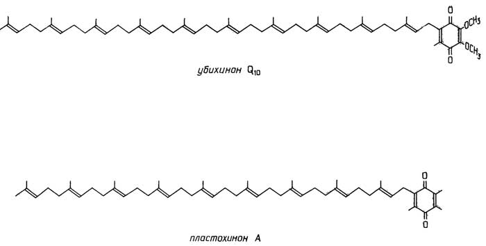
Fig. II.85. Terpenoid hydrogen carriers in oxidation-reduction reactions
of sharks as well as in human skin sebum. A triterpene chain is part of vitamin K2.
Tetraterpenes (Fig. II.84) are primarily various colored pigments. Some of them accumulate in storage tissues of plants (such as tomatoes and carrots), while others participate in light-harvesting processes in many photosynthetic organisms.
Terpenes with a chain length greater than eight isoprene units (Fig. II.85). Ubiquinones (coenzymes Q) participate in oxidation-reduction reactions occurring in bacteria and mitochondria. The most widespread is ubiquinone with a pentaterpene chain, coenzyme Q10, though coenzyme Q8 and coenzyme Q6 are also known. Plastoquinones are analogs of ubiquinones but occur exclusively in the chloroplasts of plant cells. The most common is plastoquinone A with a terpene chain consisting of 9 isoprene units, although plastoquinones with 6, 8, and 10 isoprene units are also known.
Polyterpenes: rubber and gutta-percha. The chains of the former consist of isoprene units with double bonds in the cis-configuration, whereas the chains of the latter contain isoprene units with double bonds in the trans-configuration.
Steroids
Steroids can be viewed as polycyclic derivatives of the triterpene squalene. The chain of the latter folds to form the steroid skeleton of perhydrocyclopentanophenanthrene (Fig. II.86). Subsequent enzymatic modification of this skeleton leads to the formation of various steroids.
General features of many steroids:
rigidity of the structure, which consists of three fused "chair" conformations (Fig. II.87);
presence of substituents—a hydroxyl or keto group at carbon C3; methyl groups at carbons C4, C10, C13, and C14;
presence of aliphatic "tails" of varying length and structure attached to carbon C17;
presence of one or more double bonds in rings A and B.
Substituents in the rings, like hydrogen atoms themselves, can be axial or equatorial. Regardless of this, a substituent (or hydrogen atom) is considered to have an α-orientation if it is positioned further from the observer in space, and a β-orientation if it is positioned closer to the observer. Consequently, steroid molecules can exhibit conformational variants. For instance, if the axially positioned hydrogen atom at C5 and the methyl group at C10 both have a β-orientation, the steroid molecule adopts a normal (cis-) conformation. If the hydrogen atom at C5 has an α-orientation and the methyl group at C10 has a β-orientation, the steroid molecule adopts an allo- (trans-) conformation. When drawing planar structural molecules, it is conventional to connect α-substituents to the steroid skeleton with dashed lines and β-substituents with solid lines.
The classification of steroids is related to the nature and number of substituents, as well as the presence of double

Fig. II.86. Structural relationship between terpenes and steroids
bonds in the rings. Steroid compounds are found in animal and plant cells, protists, and particularly fungi. In prokaryotes, steroids are extremely rare.
Sterins (sterols) are steroids characterized by an equatorially positioned, β-oriented hydroxyl group at carbon C3 and an 8–10 carbon hydrocarbon chain at carbon C17: cholesterol, phytosterols, ergosterol.
Cholesterol is the best-known sterol, widely distributed in animal tissues and to a lesser extent in plant tissues. In the animal liver, it is synthesized from acetate through the formation of several dozen intermediates, among which, alongside squalene, lanosterol and 7-dehydrocholesterol should be noted (see Fig. II.86). The former is a component of lanolin—sheep wool fat—while the latter accumulates in the skin, where it is converted into vitamin D under ultraviolet irradiation. In the animal body, cholesterol itself serves as a precursor for many other steroids.

Fig. II.87. Conformation of the steroid skeleton. Only carbon atoms are numbered
Phytosterols, or plant tissue sterols (Fig. II.96), such as methylenecycloarthenol, campesterol, sitosterol, and stigmasterol, differ from animal sterols—such as lanosterol or 7-dehydrocholesterol, known as zoosterols—by the presence of additional carbon atoms in the aliphatic tail attached to the C17 atom of the steroid skeleton. The biosynthetic pathways of phytosterols and zoosterols coincide in their initial stages (up to the formation of squalene and its 2,3-epoxy derivative) and then diverge: lanosterol is formed further in animal tissues, and cycloarthenol in plant tissues (Fig. II.96).
Ergosterol (Fig. II.96) is a mycosterol, as it is synthesized in fungi.
Sterides are esters of sterols and higher fatty acids. For instance, cholesterol is present in blood plasma in two forms: free (35%) and as cholesteryl ester (65%), containing certain fatty acids, primarily unsaturated ones. Since
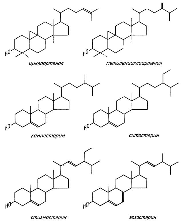
Fig. II.88. Plant sterols (phytosterols) and fungal sterols (mycosterol)
both forms are in dynamic equilibrium, cholesterol thus serves as a carrier of fatty acids. Lanosterol exists in free form as an intermediate in cholesterol biosynthesis, whereas in sheep wool fat it appears

Fig. II.89. Bile acids
primarily as an ester of myristic, arachidonic, and other rarer higher fatty acids.
Bile acids (Fig. II.89) are steroids whose common feature is the presence of an equatorially positioned and α-oriented hydroxyl group at the C3 atom and an equatorially positioned and β-oriented five-carbon hydrocarbon "tail" with a terminal carboxyl group at the C17 atom. The hydroxyl groups at the C7 and C12 atoms are also α-oriented but positioned axially. Among the four acids isolated from human bile, cholic acid predominates, but all four are synthesized in the liver from cholesterol and accumulate in the gallbladder as conjugates with glycine or the aminosulfonic acid taurine, which ensures their high water solubility. The molecular structure of all four acids adopts an A/B-cis normal conformation.
The functional role of bile acids is the emulsification of lipids in the small intestine, without which the digestion of the latter is impossible.
Human adrenal cortex hormones (Fig. II.90) are characterized by the presence of a keto group at the C3 position, an axially positioned β-hydroxyl group at the C11 atom, and a double bond between the C4 and C5 atoms. Their side chain at the C17 atom is only two carbon atoms long. Corticosterone, cortisol (hydrocortisone), and aldosterone are the three main hormones of the adrenal cortex. Cortisol enhances the conversion of amino acids into glycogen and slows down protein synthesis, whereas aldosterone regulates the water-salt balance in body tissues. Corticosterone exhibits the properties of both, though to a lesser extent. In addition, cortisol and its analogues (see Fig. II.90), such as cortisone (11-deoxycortisol) or synthetic prednisolone, are well known for their potent anti-inflammatory, anti-allergic, and immunosuppressive effects.
Progestins are hormones of the corpus luteum, placenta, ovaries, and adrenal glands. The principal hormone among them is progesterone. They ensure the normal course of pregnancy and participate in the regulation of the menstrual cycle. Progesterone and its synthetic analogues (Fig. II.91) are widely used in medicine.
Sex hormones (Fig. II.92)—male androgens and female estrogens—differ by the absence of a hydrocarbon tail at the C17 position and the retention of a keto group or a hydroxyl group at the C3 atom. Estrogens differ significantly from androgens in that ring A of estrogens is aromatic, which imparts acidic properties to their hydroxyl group. Androgens and estrogens are synthesized in the testes, ovaries, and in small amounts as intermediates in the adrenal cortex. In the male body, the ratio of androgens to
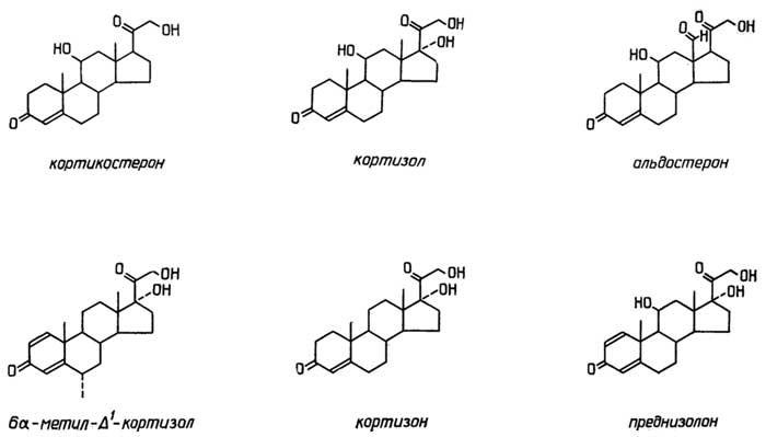
Fig. II.90. Adrenal cortex hormones and some of their synthetic analogues
estrogens is heavily shifted toward the former, and vice versa in the female body. Among male sex hormones, testosterone exhibits the highest activity, while estradiol is the most active among female sex hormones. Sex hormones regulate the functioning of reproductive
|
|
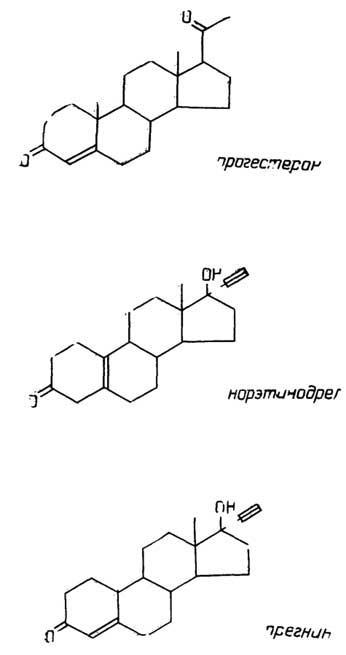

organs. The ratio of sex hormones determines the expression of secondary sexual characteristics in the body. Furthermore, sex hormones exert specific effects on other tissues and organs as well. For instance, androgens possess pronounced anabolic activity, i.e., they promote the development of skeletal muscles and their
strength. In animal husbandry and sports, synthetic anabolic agents with reduced androgenic activity are utilized (see Fig. II.92).
Vitamin D refers to two steroids designated as vitamins D2 and D3 (Fig. II.93). Vitamin D1 is an artifact; yeast ergosterol was mistakenly identified as this vitamin when, upon irradiation

Fig. II.93. Vitamin D
with ultraviolet light, it was converted into vitamin D2 (ergocalciferol). Vitamin D3 (cholecalciferol) enters the human body with food or is synthesized within it—also under the action of ultraviolet light—from 7-dehydrocholesterol accumulating in the skin. Subsequently, vitamins D2 and D3 undergo hydroxylation first in the liver (at the C25 atom) and then in the kidneys (at the C1 and C24 atoms), being converted into the active form (see Fig. II.93), which directly participates in the regulation of bone tissue mineralization.

Fig. II.94. Examples of steroid toxins
Steroid toxins (Fig. II.94) find application in pharmacology as anti-feedant (anti-attractant) substances. In particular, natural glycosidic derivatives of some of these compounds (cardiac glycosides) are used as therapeutic agents to stimulate heart function.

Saponins are substances whose molecules consist of a sugar moiety and a plant-derived aglycone with a steroid structure.
Steroid hormones are found in plants and insects. The former include gibberellins (Fig. II.95), which regulate plant morphology; about 40 of them are known. They are synthesized from diterpene through multiple stages. The latter include ecdysones, which regulate insect molting. Certain plants also synthesize them, evidently as a defense mechanism against insects.
Steroid antibiotics are rare. An example is fusidic acid (Fig. II.96), which selectively inhibits the growth of Gram-positive staphylococci.
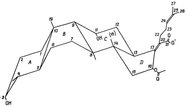
Fig. II.96. Steroid antibiotic — fusidic acid
§ 3.
Biologically Active Compounds
Related to Lipids:
Derivatives of Arachidonic Acid
The compounds listed below are synthesized within cellular organisms from arachidonic acid, whose precursor, in turn, is linoleic acid. Linoleic acid is not synthesized in the animal body and must be obtained from the diet.
Prostaglandins (Fig. II.97) are C20 carboxylic acids featuring a cyclopentane ring (C3–C12) within their chain. The letter designation of a prostaglandin (A, B, E, F, C, H) is determined by the nature of the substituents in the chain (at atoms C9 and C11), while the accompanying number indicates the number of double bonds in the chain (one at atom C13, two at C5 and C13, three at C5, C13, and C17). All prostaglandins possess a hydroxyl group at the C15 atom. Prostaglandins act as regulators (modulators) of hormone action, functioning at extremely low physiological concentrations (on the order of 10-10 mol/L). They are not produced by any endocrine glands, but are instead synthesized by cells across a wide variety of tissues. The physiological effect of the same prostaglandin can vary depending on the tissue. Prostaglandins induce the contraction or relaxation of smooth muscle,
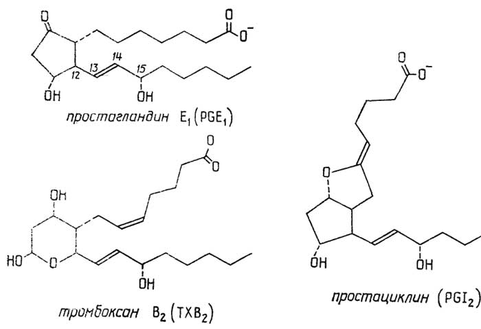
Fig. II.97. Prostaglandins
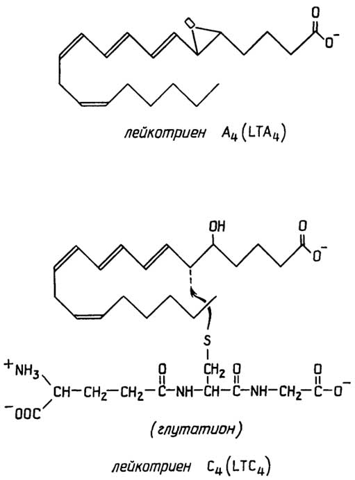
Fig. II.98. Leukotrienes
and influence blood pressure, cardiac muscle activity, and the endocrine system. They play a key role in the body's inflammatory and allergic responses, affect ion transport across certain membranes, and control synaptic nerve impulse transmission.
Thromboxanes and prostacyclins are a specialized subclass of prostaglandins that affect blood clotting.
Leukotrienes (Fig. II.98) are C20 carboxylic acids synthesized in leukocytes, sharing a common structural feature: three conjugated double bonds (at atoms C7, C9, and C11). Their physiological effect manifests as strong smooth muscle contraction in certain organs, leading to the narrowing of their lumen—such as the constriction of blood vessels in specific situations or bronchioles during asthma attacks. The active form of leukotrienes consists of their conjugates (via S) with glutathione or cysteine.
Chapter 14
LIPID COMPOUNDS
WITH SUBSTANCES OF OTHER CLASSES
AND LIPID-LIPID ASSOCIATES
§ 4.
Molecular Complexes of Lipids
with Sugars and Proteins
Glycophospholipids (Fig. II.99) are lipids whose molecules are composed of components A, B, C, and E, where component B is glycerol, and component E includes simple carbohydrates (glycerol, inositol) and sugars. In the latter case, glycophospholipids are referred to as phosphatidyl sugars. Glycerol acting as component E can, in turn, attach an amino acid to form a lipoamino acid, or serve as a bridge between two phosphatidic acid residues, forming the well-known cardiolipin (Fig. II.108).
Glycolipids, unlike glycophospholipids, lack component C (i.e., orthophosphate), and their component B can be either glycerol (glycosyldiacylglycerols) or sphingo- and dihydrosphingosine (glycosphingolipids).
Glycosyldiacylglycerols (Fig. II.101) are compounds composed of components A, B, and E, with component B being glycerol. They have been isolated from plant tissues and bacteria. Component E typically consists of a monosaccharide or disaccharide molecule, which may be partially esterified with sulfate.
Glycosphingolipids (Fig. II.102) are compounds of the composition ABE, in which component B is sphingosine (or dihydrosphingosine). They represent sugar-ceramide conjugates:
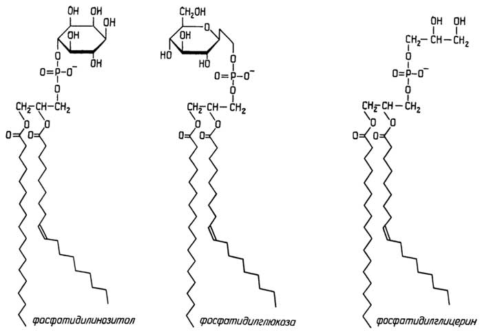
Fig. II.99. Glycophospholipids
cerebrosides are compounds of a ceramide with a monosaccharide, usually β-D-galactose or β-D-glucose, in which the hydroxyl group of the C3 atom may be esterified with sulfate;
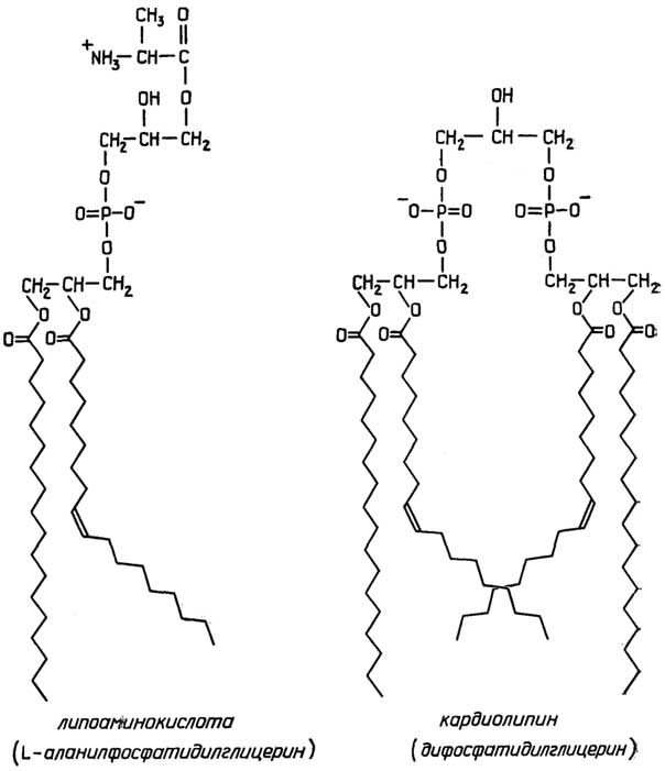
Fig. II.100. Important derivatives of phosphatidylglycerol
ceramide oligosaccharides are compounds of a ceramide with an oligosaccharide;
gangliosides are ceramide oligosaccharides containing a residue of N-acetylneuraminic acid in their carbohydrate chain. Gangliosides are crucial components of the outer leaflet of cell membranes. They largely determine cell surface specificity and the reception of environmental signals, thereby functioning as cellular antigens.
Lipopolysaccharides are highly complex polymers (Fig. II.103). Their core is based on a repeating unit of two N-acetylglucosamines linked by a β(1→6) glycosidic bond and a pyrophosphate group at the anomeric carbon atom. Two N-acyl groups represent residues of 3-D-oxymyristic
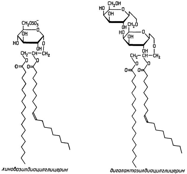
Fig. II.101.Glycosyldiacylglycerols
acids. Of the four remaining hydroxyl groups of the disaccharide, three are esterified with palmitic, lauric, and 3-D-oxymyristic acids (the latter, in turn, is esterified at its hydroxyl group with myristic acid). Each repeating unit has six fatty acid hydrophobic tails directed downward. This part of the lipopolysaccharide is designated as lipid A. The fourth hydroxyl group of the disaccharide is linked to a branched hydrophilic oligosaccharide, commonly referred to as the lipopolysaccharide core oligosaccharide. Extending further from the core oligosaccharide is the polysaccharide chain,
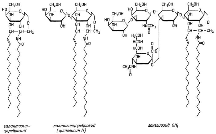
Fig. II.102.Glycosphingolipids

Fig. II.103.Lipopolysaccharide from the cell wall of the Gram-negative bacterium Salmonella typhimurium (serotypes A and B)
which is built from a tetrasaccharide repeating unit dozens of times over. It is these O-side chains that exhibit antigenic properties, which is why they are named O-antigens.
Proteolipids are globular proteins characterized by three distinct features:
a significant amount of hydrophobic amino acids within the polypeptide chain;
a substantial number of fatty acids attached to the polypeptide chain via ester bonds;
the ability to function only in a lipid environment.
Proteolipids are a type of membrane proteins (specifically enzymes) that are widely distributed in nature. They are insoluble in water but can be solubilized in organic solvents.
§ 5.
Special Properties of Lipids:
The Ability to Form Aggregates
Unlike proteins, nucleic acids, or polysaccharides, lipids are not polymers; however, due to the specific features of their molecular structure, they can form large aggregates under natural conditions that resemble macromolecular substances. Low-polarity lipids, such as neutral fats and energy-storing waxes, form aggregates in the form of oil droplets. The amphiphilicity (the property of molecules possessing both a hydrophilic and a hydrophobic part) of structural lipids determines their ability to form aggregates, whose shape and size depend on the nature of the lipid, its concentration, as well as the composition and polarity of the medium.
The types of aggregates formed by amphiphilic substances (also known as substances with amphipathic properties) can be as follows (Fig. II.104):
micelle in the form of a sphere or ellipsoid;
monolayer at the interface between two immiscible solvents of different polarity (with different dielectric constants);
bilayer, which also exists in the form of multi-layered lamellar structures, liposomes, and vesicles;
structure with hexagonal packing.
The formation of aggregates itself begins when the concentration of the amphiphilic substance in the solvent reaches a certain threshold known as the critical micelle concentration (CMC).
Natural lipids with amphipathic properties, such as phospholipids or glycolipids, tend to form bilayer-structured aggregates. Micelle formation is possible, but only at very low lipid concentrations (CMC ≈ 10-10 mol/L); furthermore, the concentration of micelles is extremely low, and the micelles themselves are quite large (the aggregation number reaches values of 2000–3000 lipid molecules).
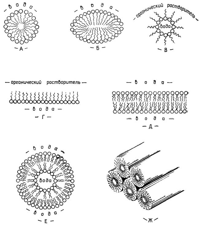
Fig. II.104.Types of aggregates formed by amphiphilic molecules:
A, B, C - micelles; D - monolayer; E - bilayer; F - vesicle (liposome - multilamellar vesicle); G - hexagonal packing
Substances with amphipathic properties, such as artificially synthesized detergents frequently used in laboratories (Fig. II.105), preferentially form micellar aggregates, with the CMC of such detergents lying within the range of 10-3–10-5 mol/L, and the number of aggregated molecules ranging from several tens to about a hundred and a half or slightly more.
The propensity of phospholipids and glycolipids to form bilayers is due to the specific feature of the hydrophobic part of their molecules—namely, the presence of two fatty acid tails, which prevents such molecules from aggregating into structures with a small radius of curvature.

Fig. II.105.Typical laboratory detergents:
A - anionic sodium dodecyl sulfate; B - cationic cetyltrimethylammonium bromide; C - neutral detergent from the Triton X group
A special position is occupied by natural steroid-structure detergents (bile acids, cholesterol), which is undoubtedly related to their structure. They form aggregates (CMC ≈ 10-3 mol/L) consisting of just a few molecules and are completely incapable of forming bilayers. However, these detergents easily incorporate into micelles or bilayers formed by other amphiphilic substances.
§ 6.
Lipoproteins and Chylomicrons:
Lipid-Protein Aggregates
Lipoproteinsand their subsetchylomicronsare molecular complexes of various lipids and proteins normally present in blood plasma. They are spherical micellar structures whose surface consists of polar phospholipid head groups and protein domains enriched in polar and charged amino acids. Beneath this hydrophilic surface lie the hydrophobic tails of phospholipids and proteins, as well as neutral fats, cholesterol, and cholesterol esters.
Based on their density, lipoproteins are conventionally classified into chylomicrons and very-low-density lipoproteins (VLDL, or pre-β-lipoproteins), low-density lipoproteins (LDL, or β-lipoproteins), and high-density lipoproteins (HDL, or α-lipoproteins). As particle density increases, particle size decreases. The density of lipoproteins rises with a higher protein content. For instance, proteins account for less than 2% of chylomicrons and, together with a small amount of phospholipids (<1%), form the chylomicron shell, which encloses triacylglycerols (over 95%) and trace amounts of cholesterol (<2%). In contrast, the protein fraction in HDL reaches 50%, phospholipids account for 30%, and the proportion of triacylglycerols drops to 3%. The highest cholesterol content is found in HDL (about 50%), whereas in VLDL and LDL it is 2.5–3 times lower. Chylomicrons are synthesized by intestinal mucosal cells and transport triacylglycerols to the liver and adipose tissue. Lipoproteins are produced by liver cells: VLDL deliver liver-synthesized triacylglycerols to adipose tissue, LDL transport cholesterol to various tissues, and HDL shuttle it back to the liver.
§ 7.
Biomembranes:
Complex Supramolecular
Lipid-Based Structures
Biomembranesare film-like structures with a thickness of 6-10 nm, characterized by a similar composition and architecture, and serving two primary functions:
to delimit the cell from its surrounding environment or compartmentalize the cell into distinct spaces known ascompartments, thereby forming various cellular organelles;
to mediate interactions and the exchange of matter between the cell and the extracellular environment, as well as between organelles and the intracellular milieu.
Biomembranes are represented (Fig. II.106) by a lipid bilayer in which proteins, glycoproteins, and glycolipids are embedded. All components are held together by non-covalent (predominantly hydrophobic) cooperative interactions. The lipid-to-protein ratio in biomembranes varies depending on their origin, ranging from 5 : 1 to 1 : 4. Protein molecules may reside on either surface of the bilayer (peripheral proteins), be partially embedded in the hydrophobic core, or span the entire bilayer (integral proteins). Glycoproteins and glycolipids constitute anywhere from a fraction of a percent to several percent of membrane components. They are invariably located on the outer leaflet of the bilayer, with their carbohydrate moieties protruding outward. The lipid matrix consists primarily of phospholipids (up to 90%), with the outer and inner leaflets differing in composition. For example, in the well-studied erythrocyte membrane, the outer leaflet contains predominantly phosphatidylcholine and sphingomyelin, whereas the inner leaflet is enriched in phosphatidylethanolamine and phosphatidylserine. The bilayer may also contain minor amounts of neutral fats—such as tri-, di-, and monoacylglycerols—as well as cholesterol and its esters, with cholesterol found primarily in the plasma membranes of animal cells.
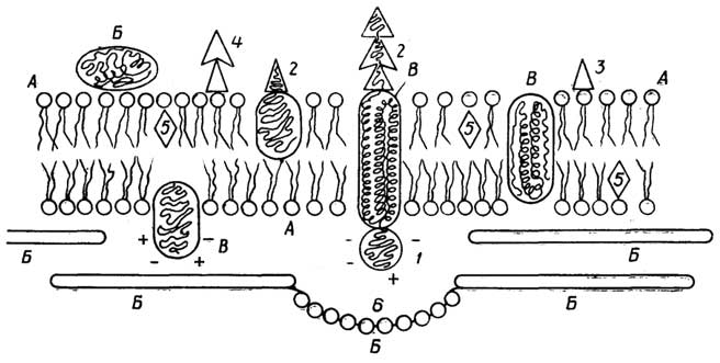
Fig. II.106.Schematic representation of an animal cell plasma membrane:
A- phospholipids;B- peripheral proteins;C- integral proteins;1- polar region of the protein globule;2- carbohydrate moieties of the protein globule;3- glycolipid;4- ganglioside;5- cholesterol;6- actin
Biomembranes of living cells possess a liquid-crystalline structure. Lipids and certain proteins move readily within the plane of the membrane. The viscosity of the bilayer is comparable to that of olive oil. The lateral diffusion of other proteins is restricted, which can be attributed either to the formation of more ordered,cluster-like structures by surrounding lipid molecules or to the protein being "anchored" to the adjacent cytoskeletal layer, which forms a resilient underlying scaffold that acts as a membrane skeleton.
Factors influencing the liquid-crystalline state and function of biomembranes include: 1) temperature; 2) ionic strength; 3) the ratio of long-chain to short-chain hydrocarbon moieties in lipids; 4) the ratio of saturated to unsaturated (with acis-configuration) hydrocarbon chains in lipids; and 5) the presence of cholesterol within the biomembrane.
§ 8.
Structural Features of the Outer Membrane
in the Cell Wall of Gram-Negative Bacteria
The primary distinction between the cell walls of Gram-negative and Gram-positive bacteria is the presence of a second (outer) membrane in the former, which overlies the rigid peptidoglycan cell wall. The outer membrane differs from the plasma membrane in its lipid and protein composition; its defining feature, however, is the presence of lipopolysaccharides on its outer leaflet. Conversely, the inner leaflet of the bilayer consists, as usual, of phospholipids and embedded proteins—specifically,envelope proteins—through which the outer membrane anchors to the murein layer of the cell wall.
Section C
NUCLEOTIDES
AND NUCLEIC ACIDS
Chapter 15
NUCLEOTIDES
Nucleotidesare compounds composed of a nitrogenous heterocyclic base, a carbohydrate (typically a pentose), and one, two, or three phosphoric acid residues.Nucleosidesare derived from nucleotides by the removal of the phosphoric acid residues. All naturally occurring nucleotides can be broadly divided into two major groups:
mononucleotides—the monomeric building blocks of nucleic acids;
other nucleotides that play an equally vital role in living metabolism.
§ 1.
Mononucleotides:
Components of Nucleic Acids
Nucleotides found in nucleic acids contain various derivatives of two heterocyclic nitrogenous bases:pyrimidine(specifically, 2-hydroxypyrimidine) andpurine(Fig. II.107).
Principal pyrimidinebases (designated by their initial letter in Latin or Cyrillic):cytosine(C or Ц),uracil, andthymine.
Principal purinebases:adenineandguanine.
Using2-hydroxypyrimidineas an example, Figure II.107 illustrates an important property of such bases: the existence of two tautomeric forms,lactimandlactam. The latter form predominates at pH 7. A lactim-lactam equilibrium also exists in other derivatives of pyrimidine and purine.
In addition to the fiveprincipalnitrogenous bases, nucleic acids contain:
Minor pyrimidinebases (Fig. II.108) are either substituted at the C5 position, such as 5-methylcytosine (m5C), 5-hydroxymethylcytosine, 5-hydroxymethyluracil, or "unusual" bases, such as dihydrouracil (D or hU) and 4-thiouracil (4s, s4U or 4S);
Minor purine bases (Fig. II.109) are various purine bases methylated at the N1 or C2 position, for example, 1-methylguanine (m1C) and 2-methyladenine (m2A), as well as those methylated or dimethylated at the amino group located at position 6 or 2, such as 6-methyl- (or 6,6-dimethyl)-aminopurine (m6A or m26A) and 2-methyl- (or 2,2-dimethyl)-amino-6-oxypurine (m2G or m22G). In so-called transport
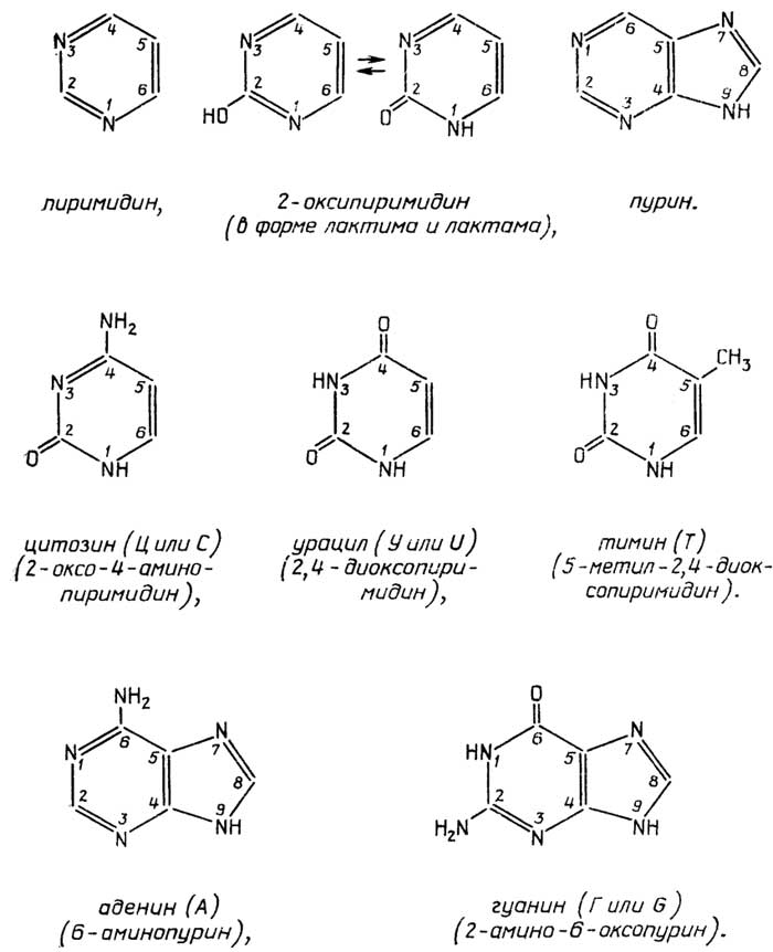
Fig. II.107. Principal pyrimidine and purine bases
ribonucleic acids (tRNAs), nucleotides containing hypoxanthine (6-oxypurine) have been found, as well as those with other 6-aminopurine derivatives featuring a more complex modified amino group.
All ribonucleic acids (RNAs) contain β-D-ribose as their sugar component, whereas deoxyribonucleic acids (DNAs) contain 2-deoxy-β-D-ribose.

Fig. II.108. Minor pyrimidine bases
The linkage of the three components in a mononucleotide is established via a bond between the N1 atom of the pyrimidine base or the N9 atom of the purine base and the C1' atom of the pentose5, and through the formation of an ester bond between the C5' atom of the pentose and phosphoric acid, as shown in Fig. II.110 for a mononucleotide formed by cytosine, deoxyribose, and phosphoric acid, referred to as 5'-deoxycytidylic acid. Its anion is deoxycytidine-5'-phosphate, or dCMP (dCMP) for short.
The relative spatial arrangement of the nitrogenous base and the pentose in a nucleoside or nucleotide can vary. In general, it is accepted that if atoms 2 and 3 of the pyrimidine

Fig. II.109. Minor purine bases
ring or atoms 1 and 2 of the purine ring lie on one side of the C1'-N glycosidic bond, while the pentose ring lies on the other, we are dealing with the anti-conformer; if the specified base atoms and the pentose ring are on the same side of the glycosidic bond—that is, the base is positioned directly above the sugar—then this is the syn-conformer of the nucleoside or nucleotide (Fig. II.111).

Fig. II.110. Formation of a mononucleotide
The principal mononucleotides entering into the composition of RNA are listed in Table II.4, and those of DNA in Table II.5.
It should be noted that dUMP is typically absent in DNA. Instead, dTMP is present, which is often incorrectly referred to simply as thymidine phosphate. dUMP is found only in the DNA of certain bacteriophages, whereas thymidine monophosphate containing ribose has been discovered in certain tRNAs. It is referred to as ribosylthymidine phosphate or ribothymidine phosphate to distinguish it from DNA thymidine phosphate.
It should not be forgotten that the aforementioned minor pyrimidine and purine bases also form mononucleotides, which occur in certain RNAs or DNAs. We should merely point out that the nucleoside containing hypoxanthine and ribose is

Fig. II.111. SYN and ANTI conformers of mononucleotides
Table II.4 Principal RNA Mononucleotides
|
Table II.5 Principal DNA Mononucleotides
|
inosine (I), and the corresponding mononucleotide is called inosinic acid or inosine phosphate (Fig. II.112). It is unrelated to inositol or inositol phosphate, despite the similarity in name (see pp. 40, 42).

Fig. II.112. Rare nucleic acid mononucleotides
Another feature to note is the unusual bond between the nitrogenous base uracil and ribose in pseudouridine-5'-phosphate (pseudouridine is abbreviated as ψ) (Fig. II.112).
§ 2.
Other Important Nucleotides
In addition to mononucleotides—the building blocks of nucleic acids—various mono- and dinucleotides occur in the cell in a free state or as components of other important compounds.
3'- and 2'-Mononucleotides
Nucleic acid nucleotides found in the cell in free form as intermediates in nucleic acid degradation and synthesis are typically nucleoside 5'-phosphates. However, 3'-phosphates can be obtained via nucleic acid hydrolysis, and in the case of RNA hydrolysis, nucleoside 2'-phosphates as well. It is standard practice to omit the pentose carbon number when referring to a nucleoside 5'-phosphate; however, mentioning the pentose carbon number is mandatory for 2'- and 3'-mononucleotides.
Cyclic Adenylic Acid (cAMP)
cAMP, i.e., adenosine-3',5'-phosphoric acid (Fig. II.113), is an important mediator of the regulatory action of hormones in numerous processes.
5'-Nucleoside Diphosphates and 5'-Triphosphates
In addition to 5'-nucleoside monophosphates, cells also contain 5'-nucleoside diphosphates and 5'-triphosphates (Fig. II.114). Their phosphate groups are designated as α, β, and γ, starting from the ester bond. They can be sequentially cleaved at the points indicated in the figure by a wavy line.
|
|

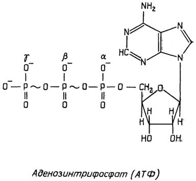
At neutral pH, 5'-nucleoside triphosphates and diphosphates, such as ATP and ADP, exist as anions with a charge of -4 and -3, respectively, since only the α-phosphate group has a pKa of 6.5–7.2; the pKa values of the remaining phosphate groups lie in the acidic region (pH around 2).
The pyrophosphate groups of ATP and ADP have a high affinity for divalent metal cations (Mg++, Mn++). ATP and ADP participate in numerous metabolic processes specifically as complexes with Mg++ (and occasionally Mn++). It is worth noting here that the toxicity of beryllium salts is due to the fact that Be++ cations also bind strongly to ATP and ADP, but such complexes act as metabolic poisons.
The special role of ATP in any living matter is determined by its primary function: serving as the principal carrier of chemical energy in the cell. As a result of catabolism—that is, the enzymatic breakdown of dietary proteins, carbohydrates, and fats—the energy of their chemical bonds is stored in all living organisms through the phosphorylation of AMP and ADP to ATP. Subsequently, via the reverse reaction
(ATP hydrolysis), this stored energy is utilized by living organisms for:
the biosynthesis of complex molecules required by the organism from simpler ones, i.e., in anabolic processes;
performing mechanical work in the form of movement or muscle contraction;
the active transport of substances against a concentration gradient;
maintaining body temperature in warm-blooded organisms.
Thus, the unique feature of the adenylate system (Fig. II.115) lies in its role as a bridge linking catabolic and anabolic processes.
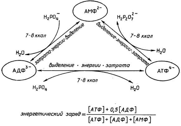
Fig. II.115. The adenylate system
Catabolism aims to increase the fraction of ATP, whereas anabolism decreases it. The ratio of the components of the adenylate system in cells is conventionally expressed as the "energy charge":
energy charge =
[АТФ] + 0,5 [АДФ] |
[АТФ] + [АДФ] + [АМФ] |
.
This formula demonstrates that the adenylate system is fully "charged," with a charge of 1, when all AMP and ADP have been converted into ATP. Conversely, if only AMP is present, the system's charge is zero. ADP represents a "semi-charged" state. Typically, the energy charge in cells ranges between 0.75 and 0.90.
Changes in the standard Gibbs free energy ΔG°(37°C) (where the prime denotes that ΔG° corresponds to pH 7, as customary in biochemistry) for the interconversions of the adenylate system components at 37°C, μ = 0.2 M, and in the presence of excess Mg++ fall within the range of 7–8 kcal. These data were obtained for standard conditions

Fig. II.116. Mononucleotides as molecule carriers
(where the concentrations of initial reactants and products are 1 M each) with adjustments for the temperature and pH prevailing within the cell. However, when accounting for actual intracellular concentrations, ΔG for ATP hydrolysis, for example, becomes a more negative value (-12.5 kcal). To determine the feasibility of a spontaneous reaction
(in the presence of a suitable enzyme catalyst), biochemistry relies on ΔG°(25°C) or ΔG°'(37°C). Because hydrolysis reactions of these compounds involve the transfer of a specific group to water, ΔG°' in such cases is referred to as the group transfer potential.
Aside from ATP, other nucleotides also participate as chemical energy carriers in biosynthetic processes:
ATP, GTP in protein biosynthesis,
ATP, UTP in polysaccharide biosynthesis;
ATP, CTP in lipid biosynthesis;
ATP, GTP, UTP, CTP in RNA biosynthesis;
dATP, dGTP, dTTP, dCTP in DNA biosynthesis.
All nucleoside 5'-triphosphates readily exchange their γ-phosphate group via nucleoside 5'-diphosphates with the assistance of the enzyme nucleoside diphosphate kinase:
ATP + UDP ↔ ADP + UTP;
ATP + GDP ↔ ADP + GTP;
ATP + CDP ↔ ADP + CTP;
GTP + UDP ↔ GDP + UTP;
ATP + dCDP ↔ ADP + dCTP etc.
It should be noted that during nucleic acid biosynthesis, nucleoside 5'-triphosphates serve a dual function: they provide not only
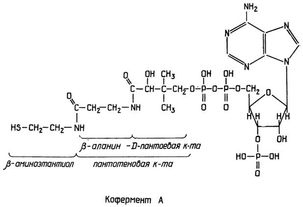
Fig. II.117. Acyl group carrier — coenzyme A
the energy required for biosynthesis to proceed, but also the building blocks themselves—mononucleotides.
Certain nucleoside 5'-phosphates act as molecular carriers. For instance, in glycogen synthesis, glucose is transferred via uridine diphosphate, while in lecithin synthesis, choline is transferred via cytidine diphosphate (Fig. II.116).
The complex nucleoside 5'-diphosphate, briefly designated as coenzyme A (Fig. II.117)—consisting of consecutively linked adenosine 3'-phosphate-5'-pyrophosphate, the vitamin pantothenic acid, and β-aminoethanethiol—functions in metabolic processes as a carrier of acyl groups, which are bound by an ester linkage to its terminal SH group.
NAD+ and NADP+
Aside from the aforementioned nucleotides, the bridge between catabolic and anabolic processes is formed by two

Fig. II.118. Mediators of oxidation-reduction reactions NAD and NADP
dinucleotides: nicotinamide adenine dinucleotide (NAD+) and nicotinamide adenine dinucleotide phosphate (NADP+) (Fig. II.118). Their linking role stems from their ability to exist in two forms: oxidized (NAD+ and NADP+) and reduced (NADH and NADPH):
NAD+/NADH |
E°' = — 0.320 V; |
NADP+/NADPH |
E°' = — 0.324 V |
(the prime here indicates that E° was determined at pH 7). Hydrogen is likely transferred in the form of a hydride ion (see Fig. II.118). A structural component of NAD+ and NADP+—nicotinamide or its precursor nicotinic acid (niacin)—is also known as vitamin PP. During the oxidation of "biological fuel" in

Fig. II.119. Mediators of oxidation-reduction reactions FMN and FAD
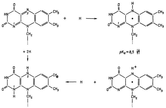
Fig. II.120. Oxidation-reduction transitions in FMN and FAD
catabolic processes, hydrogen is "stored" within NADH and NADPH molecules. Hydrogen is subsequently removed from these molecules during anabolism and utilized for hydrogenation in the biosynthesis of compounds essential to cell survival. NAD+ is predominantly employed in catabolic processes, whereas NADPH is more widely involved in anabolic reactions. Hydrogen exchange between the two carriers according to the scheme
NADH + NADP+ ↔ NAD+ + NADPH
readily occurs with the aid of enzymes known as transhydrogenases.
Two other important hydrogen carriers are flavin mononucleotide (FMN) and flavin adenine dinucleotide (FAD) (Fig. II.119). A constituent part of FMN and FAD is riboflavin, commonly known as vitamin B2. It represents a compound composed of a complex nitrogenous base, isoalloxazine, and a five-carbon sugar, ribitol. The reversible redox transition
FMN ↔ FMNH2 or FAD ↔ FADH2
is associated with the addition and removal of two hydrogen atoms at the N1 and N5 positions of isoalloxazine, with the potential formation of a semireduced form—the semiquinone radical (Fig. II.120). The formation of the reduced forms FMNH2 and FADH2 can also be explained by the addition and removal of a hydride ion and a proton at the N5 and N1 positions, respectively.
5 Unlike the atoms of the nitrogenous base, pentose atoms are designated with a prime.
Chapter 16
NUCLEIC ACIDS
§ 1.
Primary Structure
Formation of the Polymer Chain
Mononucleotides are linked into polymers—natural or synthetic nucleic acids (NAs)—via the formation of ester bonds between the 3'-carbon of one mononucleotide pentose, a phosphate group, and the 5'-carbon of another mononucleotide pentose (Fig. II.121).
When writing out the primary structure in an abbreviated form, the 5'-end of the chain is placed on the left and the 3'-end on the right, for example
5'-endpApCpGpUpm2Apψpm5Cpm22Gps4UpC3'-end. The number of nucleotide units in RNA ranges from several dozen in the shortest RNA molecules to 2 · 105 in the longest. RNAs generally contain significantly more nucleotides with minor bases than DNAs. The shortest DNAs consist of several thousand units, while the largest contain up to 108 units. The length of such chains can reach several centimeters.
Features of the Primary Structure of Nucleic Acids
RNAs typically consist of a single polymer chain, whereas DNAs, by contrast, most commonly form a two-chain structure held together by non-covalent interactions. These chains are always antiparallel, meaning that the 5'-end of one chain lies adjacent to the 3'-end of the other.

Fig. II.121.Formation of a nucleic acid polymer
However, double-stranded RNAs and single-stranded DNAs are also known. The covalent chains of natural polynucleotides can be strictly linear, branched, or cyclized, and these rings may be intertwined (interlocked with one another). Some single-stranded nucleic acids (or artificially synthesized polynucleotides) can fold back on themselves in certain regions, thereby forming double-stranded "hairpin-loop" structures. Even triple-stranded polynucleotides can be obtained artificially.
Hydrogen bond formation between
bases is the primary driver for the
association of polynucleotide chains
Chargaff's rules for double-stranded DNA state that the mole fraction of adenine equals the mole fraction of thymine, and the mole fraction of guanine equals the mole fraction of cytosine; that is, m.f. A = m.f. T, and m.f. G = m.f. C, or alternatively, (1 - m.f. AT) = m.f. GC. The values of m.f. GC range from a minimum of 0.005 to a maximum of 0.75. When calculating the mole fraction of each major base, all of its derivative minor bases are taken into account. Chargaff's rules for double-stranded RNAs (such as those of certain viruses) hold true provided that uracil (U) and its derivatives are substituted for thymine (T). The validity of Chargaff's rules for double-stranded DNA and RNA structures enabled Watson and Crick to propose a model for nucleic acid structure based on the complementary pairing of adenine with thymine (or uracil) and guanine with cytosine through hydrogen bonding (Fig. II.122). The distance between the C1 atoms of the pentoses from opposing chains is 1.085 nm in both cases, with the AT pair held together by two hydrogen bonds and the GC pair by three.
It is worth noting that other atoms within the bases are also capable of forming hydrogen bonds. Consequently, an alternative mode of base interaction can be envisioned (Fig. II.123)—known as the Hoogsteen or imidazole scheme. In this type of interaction, the distance between the pentose C1 atoms is shorter than in the Watson-Crick model, measuring 0.88 nm. This interaction motif has been observed in the co-crystallization of methylated derivatives of adenine and thymine, though it does not occur within the standard double helix.
§ 2.
Secondary Structure
DNA
Cellular DNA is double-stranded. Single-stranded DNA has been found in certain viruses.
A key characteristic of DNA is that its composition and structure are identical across all cells of a given organism, as well as in every virion of a given viral species, and remain unchanged throughout the organism's lifetime or under varying environmental conditions6. Its nucleotide composition and architecture serve as
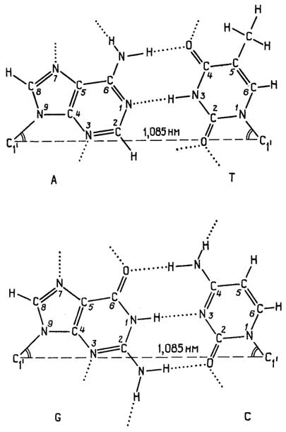
Fig. II.122.Formation of hydrogen bonds between complementary nitrogenous bases according to Watson and Crick (direct scheme)

Fig. II.123.Formation of hydrogen bonds between complementary nitrogenous bases according to Hoogsteen (direct scheme)
a defining characteristic of the organism. This is because the nucleotide sequence of DNA encodes all the genetic information passed down from generation to generation. The composition of DNA is sometimes conventionally characterized by the ratio
m.f. (A + T) |
m.f. (C + G) |
.
According to Watson and Crick, the double-stranded DNA molecule forms a right-handed double helix. The two strands run in antiparallel directions. The chains are complementary in their nitrogenous bases and are held together primarily by hydrogen bonds. The nucleotides adopt the anti conformation. Consequently, pairs of hydrophobic nitrogenous bases stack flat upon one another inside the core of the helix, while the hydrophilic sugar-phosphate backbones wind around the outside of this stack. A second force—no less important than hydrogen bonding in stabilizing the double helix—is base stacking interactions among the stacked bases. The nature of stacking appears to involve both induced dipole interactions of π-bonds and hydrophobic effects. It should be noted, however, that unlike hydrophobic interactions in proteins, thermodynamic parameters during stacking change in the opposite direction; that is, ∆H and ∆S are negative, and ∆G is negative solely because the enthalpic contribution outweighs the entropic one.
The DNA double helix can adopt several different conformational forms (Fig. II.124): B, A, C, Z.
B-form of DNA, isolated from aqueous solutions. Its characteristics are:
helix diameter of 2.1 nm;
10 base pairs per turn, helix pitch of 3.4 nm;
base pairs are perpendicular to the longitudinal axis of the helix;
the longitudinal axis of the helix passes right through the middle of the base stack;
there are two grooves between the strands: a major groove and a minor groove.
A-form of DNA can be obtained from aqueous solutions by adding solvents with a lower dielectric constant, such as alcohol. It is characterized by:
helix diameter of 2.5 nm;
11 base pairs per turn, helix pitch of 2.8 nm;
base pairs are tilted at an angle of 20° to a plane perpendicular to the longitudinal axis of the helix;
they wind around the longitudinal axis in such a way that a cavity with a diameter of 0.8 nm runs along its center;
it also features two grooves, but the major groove is wider and deeper.
The C-form, Z-form of DNA, and several other less-studied forms are also known.
The C-form is characterized by:
9.3 base pairs per turn;
base pairs are tilted at an angle of about 6° to a plane perpendicular to the longitudinal axis of the helix;
base pairs are slightly inclined relative to each other.

Fig. II.124. Two forms of DNA
The Z-form differs significantly from the others in that:
its two strands are wound into a left-handed helix with 12 base pairs per turn;
the sugar-phosphate backbones do not form smooth lines, unlike in the B- or C-forms of DNA, but instead have a jagged, zigzag (Z) appearance.
RNA
Cellular RNAs are single-stranded. Double-stranded RNAs have been found in certain viruses.
RNA exists in several forms that differ in biological function, structure, and properties.
rRNA (ribosomal RNA) accounts for 80-90% of total cellular RNA. Its chain is intricately folded into highly compact structures stabilized by the incorporation of numerous proteins. Such complexes are insoluble under normal conditions and can be isolated by centrifugation. Although the rRNAs of bacteria, plants, and animals differ slightly, in each case they can be separated into several molecular species that differ in their sedimentation coefficient S (Table II.6). rRNA is one of the major components of the polypeptide synthesis machinery.
Table II.6
Types of rRNA in prokaryotes and eukaryotes
Designation |
Bacteria |
Plants and animals |
Number of nucleotides |
Molecular |
"Light" rRNA |
5S |
5S, 5.8S |
100-150 |
3 · 104- 5 · 104 |
pre-rRNA are precursor molecules of ribosomal RNAs. Each of the three molecular types of rRNA is formed from a longer pre-rRNA. All of them, in turn, are cleavage products of a single transcript (a complementary ribonucleic acid copy of a region of one of the DNA strands).
tRNA stands for transfer, or soluble, RNA. It accounts for up to 15% of all cellular RNA. Its chain is folded into a globule. It is readily soluble under normal conditions. The function of tRNA is to deliver amino acids to the site of polypeptide chain synthesis. Each amino acid corresponds to one or more tRNAs. There are about 100 known tRNAs in total. The number of nucleotides in a tRNA molecule reaches several dozen. The molecular weight is approximately 3 · 104.
pre-tRNA are precursor molecules of tRNA. They typically include nucleotide sequences of more than one tRNA.
mRNA stands for messenger, or informational, RNA. It has a short lifespan in the cell, ranging from a few minutes to several hours, or occasionally up to tens of hours. At any given moment, it constitutes about 3% of total cellular RNA. Single-stranded, extended mRNA molecules are polymers with a molecular weight ranging from 2.6 · 104 to 1 · 106, containing from several dozen to several thousand nucleotides. mRNA represents complementary copies of a region of one of the two DNA strands. The sequence of amino acids in the polypeptide chain is encoded in the alternation of nucleotides. Thus, each protein corresponds to its own mRNA. Sometimes, mRNA carries information for two or more proteins.
hnRNA stands for heterogeneous nuclear RNA. This is a higher-molecular-weight precursor of mRNA with an even shorter half-life. The number of nucleotide units ranges from 1500 to 3000. Its molecular weight is from 105 to 2 · 107. It has been detected exclusively in eukaryotic cells.
snRNA stands for small nuclear RNA, also found in eukaryotes. Short snRNA molecules contain between 65 and 200 nucleotides. They are essential for the processing of hnRNA into mRNA.
Viral RNA is often classified as a distinct molecular type due to its unique features. Its characteristics are that it:
like DNA, serves as a carrier of hereditary information;
unlike other RNAs, can also be double-stranded.
Viral RNA, whether single-stranded or double-stranded, is packed extremely compactly within viral particles through mechanisms that remain unclear. It contains several thousand bases or base pairs. Its molecular weight falls within the range of 106- 107.
To date, secondary structure has been thoroughly studied in tRNAs and partially determined in double-stranded viral RNAs.
The structure of certain tRNAs has been fully deciphered. There is little doubt that it is broadly similar across all tRNAs. Their secondary structure is known as the "cloverleaf" model. It is maintained by the formation of about 50 hydrogen bonds across two dozen pairs of complementary bases (Fig. II.125). Common features of all tRNAs include:
the ability to attach amino acids to the 3'-end via the formation of an ester bond between the amino acid and the 2'- or 3'-hydroxyl groups of the terminal adenosine;
the presence of a conserved A-C-C sequence at the end of the acceptor stem (3'-end);
base pairing via hydrogen bonds occurring at conserved positions;
the dihydrouridine loop (D-loop), which varies in size but shares significant sequence homology; it contains a region responsible for binding the enzyme that catalyzes the joining of tRNA-delivered amino acids into a polypeptide chain;
the pseudouridine loop (ψ-loop), which contains a large conserved region identical across all tRNAs and is responsible for binding to tRNA during protein synthesis;
some tRNAs feature a loop of varying length known as the variable loop, which contains identical nucleotides at specific conserved positions; various hypotheses exist regarding its functional role;
the anticodon loop contains a triplet of nucleotides (the anticodon) through which tRNA interacts with the corresponding triplet (the codon) of mRNA during protein biosynthesis; the anticodon and codon are complementary; the anticodon is always preceded by uridine in the loop.
The secondary structure of double-stranded viral RNAs has been investigated using X-ray diffraction analysis. Based on these data, two structural variants appear possible:
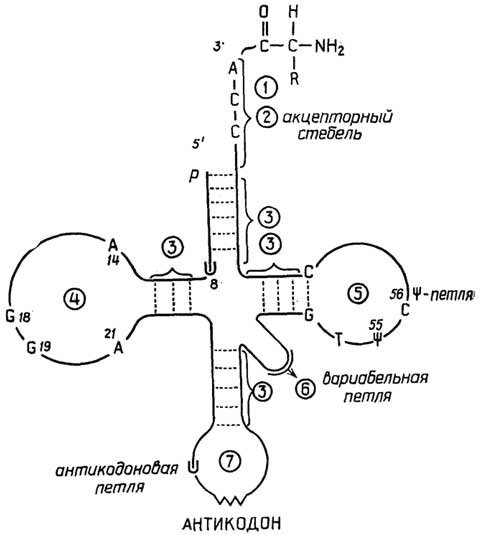
Fig. II.125. tRNA "cloverleaf" structure
double-stranded right-handed RNA-11 helix, which is characterized by 11 base pairs per turn, with the base pairs tilted at an angle of 13-14° relative to the plane perpendicular to the longitudinal axis of the helix;
double right-handed RNA-10 helix, which has 10 base pairs per turn, with the base pairs tilted at a 10° angle relative to the plane perpendicular to the longitudinal axis of the helix. Thus, RNA-11 is very similar to the A-form of DNA, whereas RNA-10 is intermediate between the A- and B-forms of DNA, though closer to the A-form than to the B-form.
Double helix
The existence of a DNA-RNA double helix is only possible in the A-form. This is because the B-form of DNA contains deoxyribose in the C2'-endo conformation, whereas the A-form of DNA, RNA-10, and
RNA-11 contain pentoses in the C3'-endo conformation. DNA transitions rather easily from one form to another; for RNA, transitioning into a conformation similar to the B-form of DNA is energetically unfavorable because the presence of the hydroxyl group at the C position creates significant steric hindrance when ribose shifts from the C3' to the C2' endo conformation.
§ 3.
Tertiary structure
The tertiary structure of nucleic acids can currently be examined only using tRNA and the supercoiling of circular double-stranded DNAs as examples.
Spatial structure of tRNA
To date, the tertiary structure of the yeast tRNAPhe molecule has been completely elucidated. It is an L-shaped, elongated globule. Its foundation is formed by a secondary structure resembling a "cloverleaf," folded such that the pseudouridine and dihydrouridine loops lie close to each other, while the variable loop is extended into a fairly linear segment (Fig. II.126). Four factors can be cited to explain this spatial folding:
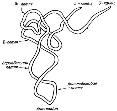
Fig. II.126. Tertiary structure of tRNA
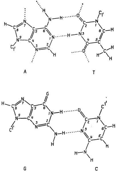
Fig. II.127. Formation of hydrogen bonds during nitrogenous base interaction via the reverse Watson-Crick scheme
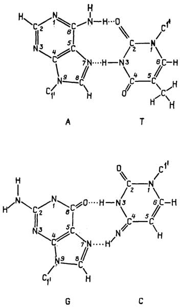
Fig. II.128. Formation of hydrogen bonds during nitrogenous base interaction via the reverse Hoogsteen scheme

Fig. II.129. Formation of hydrogen bonds among three nitrogenous bases
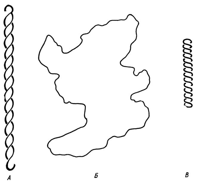
Fig. II.130. Supercoiling of circular double-stranded DNAs
the formation of hydrogen bonds between nucleotide bases located in distant regions of the "cloverleaf," such as in the D and ψ loops; this can occur via both the Watson-Crick and Hoogsteen schemes, where the plane of one of the bases in either case may be rotated by 180° (Fig. II.127; II.128);
the formation of hydrogen bonds among three bases; such triple interactions are particularly characteristic of the regions within the globule where the D, ψ, and variable loops come into contact (Fig. II.129);
the formation of hydrogen bonds between nitrogen heteroatoms, amino and oxy groups of the bases on the one hand, and phosphate groups and pentose hydroxyl groups of the RNA polymer backbone on the other; the potential for such hydrogen bonds is indicated by dashed lines in the final figure, and they are indisputable in a number of specific cases;
base stacking; only a few bases in the tRNAPhe molecule do not participate in stacking, while the rest form two base stacks that make up the two arms of the L-shape.
Supercoiling of circular
double-stranded DNAs
Although DNA molecules can reach several centimeters in length, in vivo they are usually packed so tightly that their length does not exceed several nanometers. This is achieved through supercoiling. Tertiary supercoiling of circular double-stranded DNAs is shown in Fig. II.130. Typically, double-stranded circular DNAs are wound into a right-handed superhelix. The addition of polynuclear aromatic compounds with a planar structure (Fig. II.131) to solutions of such DNAs leads to their intercalation, i.e., their insertion into the DNA base stack. An increase in the concentration of intercalating compounds causes the right-handed superhelix to unwind until it completely disappears, after which the circular double-stranded DNA molecule becomes wound into a left-handed superhelix.
§ 4.
Quaternary structure
Little is known about the quaternary structure of nucleic acids. In certain oncogenic viruses, RNA appears to consist of two identical subunits, the association between which is most likely maintained by complementary base interactions.

Fig. II.131. Examples of compounds capable of intercalating into nucleic acids and disrupting their structure
§ 5.
"External" factors
stabilizing the secondary, tertiary,
and quaternary structures of nucleic acids
In addition to base-pairing hydrogen bonds and base stacking, the stabilization of the secondary, tertiary, and quaternary structures of nucleic acids is also achieved through the "shielding" of the negative charges of phosphoric acid residues by positive charges:
metal cations such as Na+, K+, Mg++, etc.;
aliphatic amines with two or more amino groups, such as:
+H3N - (СН2)4- NH3+- putrescine,
+H3N - (СН2)5- NH3+- cadaverine,
+H3N - (СН2)4- NH2+- (СН2)3- NH3+- spermidine,
+H3N - (CH2)3- NH2+- (CH2)4- NH2+- (CH2)3- NH3+- spermine;
basic proteins—histones and protamines (small proteins rich in lysine and arginine).
6Here we exclude cases of mutation.
Chapter 17
NUCLEOPROTEINS —
SUPRAMOLECULAR COMPLEXES
OF NUCLEIC ACIDS AND PROTEINS
§ 1.
Chromosomes of Cell Nuclei
Cell nuclei contain a substance called chromatin, which consists of equal masses of DNA and basic histone proteins, as well as a small amount of acidic proteins. Histones consist of 100–200 amino acid residues and have a molecular weight of 10,000–20,000. Based on their relative lysine and arginine content, they are divided into five types: H1, H2A, H2B, H3, and H4. Remarkably, histones of the same type from completely different organisms (e.g., peas and cattle) are virtually identical in composition and structure. Under an electron microscope, chromatin appears as fibers with closely strung beads. Each bead is a nucleosome, otherwise known as a v-particle. It (Fig. II.132) constitutes
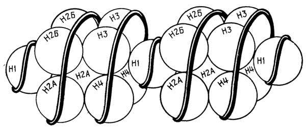
Fig. II.132. Structure of chromatin
an octamer containing two molecules each of histones H2A, H2B, H3, and H4, wrapped externally by a DNA superhelix with 150 base pairs. Between the nucleosomes, the DNA superhelix contains about 50 base pairs and is associated with only one molecule of the H1 protein. A chromosome is a chromatin fiber that is, in turn, coiled into a helix. This mechanism ensures the ultra-compact packaging of DNA.
§ 2.
Ribosomes
Ribosomes are cellular particles containing RNA and proteins, the latter accounting for less than half of their mass. A ribosome consists of two subunits—large and small—into which it dissociates if the Mg++ concentration drops below 1 mM. Each subunit is a complex of one or two RNA molecules and two to three dozen protein molecules. By gradually increasing the concentration of urea and LiCl, all RNAs can be sequentially extracted and the proteins separated. The large subunit typically contains "light" and "heavy" RNA, while the small subunit contains "medium" RNA. The scheme of the step-by-step dissociation of E. coli bacterial ribosomes is shown in Fig. II.133.

Fig. II.133. Scheme of bacterial ribosome dissociation
Polysomes (polyribosomes) are cellular structures consisting of an elongated mRNA molecule surrounded by globular proteins, onto which ribosomes are strung.
§ 3.
Informosomes
Informosomes are nucleoproteins composed of mRNA and globular proteins known as informatins, which stabilize and protect it.
Informomers are analogous nucleoproteins consisting of hnRNA and informatin proteins.
§ 4.
Viruses
Viruses represent a particularly interesting case of nucleoproteins. They are structures that exist on the borderline between the living and the non-living. On the one hand, they can be isolated as pure crystals that retain their properties indefinitely, which is characteristic of non-living matter. On the other hand, they exhibit all the properties of living things, with the caveat that they lack their own metabolic apparatus. Therefore, a virion (viral particle) can only realize its capacity for self-replication once it enters the cell of an organism (bacterium, plant, or animal) and hijacks its metabolic processes. A viral particle consists of a tightly packed single-stranded or double-stranded DNA or RNA molecule enclosed in a coat. In some cases, the coat, called a capsid, consists of a large number of protein subunits called capsomeres. In other cases, it is a membrane made of lipids and proteins (glycoproteins), in which proteins significantly outnumber lipids. The composition and structure of the virion surface determine the tissue specificity of the virus. Cell infection occurs when either the entire virion or merely its DNA or RNA penetrates the cell. Thereafter, the viral DNA or RNA reprograms the cell's metabolic processes so that ambient energy and raw materials are utilized to synthesize new viral particles.
Recommended Reading
Villee, C., and Dethier, V. Biological Principles and Processes. Moscow, 1974.
Metler, D. Biochemistry. Moscow, 1980.
White, A., Handler, P., Smith, E., et al. Principles of Biochemistry. Moscow, 1981.
Stryer, L. Biochemistry. Moscow, 1984.
Lehninger, A. Principles of Biochemistry. Moscow, 1985.
Musil J., Novaková O., Kunts K. Modern Biochemistry in Diagrams. Moscow, 1984.
Rees E., Sternberg M. From Cells to Atoms: An Illustrated Introduction to Molecular Biology. Moscow, 1988.
Ovchinnikov Yu. A. Bioorganic Chemistry. Moscow, 1987.
Berezov T. T., Korovkin B. F. Biological Chemistry. Moscow, 1983.
Filippovich Yu. B. Fundamentals of Biochemistry. Moscow, 1986.
Anisimov A. A., Leontyeva A. N., Alexandrova I. F. et al. Fundamentals of Biochemistry. Moscow, 1986.